
Mast cell tumours in dogs

What is a mast cell tumour?
Mast cells are immune cells (a type of white blood cell) that reside in many tissues throughout the body. Dogs have many of these mast cells in their skin. Mast cells contain granules, or packets of chemicals, such as histamine, serotonin and heparin. The primary function of the mast cells is to respond to inflammation and allergies by releasing these biochemicals when triggered by the immune system.
Mast cell tumours in dogs, also called MCTs, mastocytomas, or mast cell sarcomas, are the most common type of dog skin cancers, accounting for around eleven percent of skin cancer in dogs, according to a 2011 study. Mast cell tumours in dogs form when the mast cells proliferate uncontrollably and accumulate in clusters, usually in the form of skin lumps. These dog tumours often appear small and insignificant, and many are benign, but they can be very serious and even life threatening.
Mast cell tumours, or mastocytomas, release large amounts of biochemicals from the mast cells into the body, which can cause significant problems such as increased stomach acidy and gastric ulcers, internal bleeding and various allergic reactions. The effect of mast cell tumours in dogs means a decrease in the quality of life for many canine sufferers. Apart from the skin, mast cell tumours can also occur in other tissues, including the spleen, liver, bone marrow, intestines and respiratory and gastrointestinal tracts. Around fifty percent of mast cell tumours are malignant; without treatment of this particular dog cancer they will progressively worsen and result ultimately in death.
Mast cell tumour in dogs, mastocytoma, dog skin cancer, dog tumour, cancer in dogs, dog cancer lump, dog skin tumour
Source: https://blogs.webmd.com/pet-tales/2011/01/twenty-five-years-of-mast-cell-tumors.html
Symptoms of mast cell tumours
Mast cell tumours in dogs are extremely variable in their location, appearance, and growth pattern. In some dogs, the tumour or lump is not obvious or even visible, and there may be few, if any, observable symptoms to indicate that the animal has developed a mast cell tumour. Many affected dogs show no signs of illness or discomfort.
Location of mast cell tumours in dogs:
- Can appear on any part of the body, but in general:
- about 50% are found on the trunk and perineal area (anus and genitals),
- 40% on the limbs and paws (extremities)
- 10% in the head and neck region
- Can appear on the skin surface or in the tissue below the surface (subcutaneous)
- Usually a solitary isolated lump or mass
- Can appear in clusters or in multiple areas
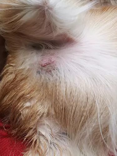
Appearance of mast cell tumours in dogs:
- Smooth, bumpy, ulcerated or rubbery
- Sore and inflamed areas on the body
- Raised round masses that feel soft on the outside and solid on the inside
- Skin mass may appear:
- Multi-nodular, i.e. several bumps clustered together, like a cauliflower
- Reddish in colour
- Hairless
- Itchy and inflamed, from high levels of histamine in the body
- Ulcerated, weeping, as an open wound
- Completely normal, just a bump under or on the skin
- May resemble other types of dog tumours or dog skin cancers
- May resemble a skin condition – insect bite, wart, allergic reaction
Growth pattern of mast cell tumours in dogs:
- Typically, they are slow-growing, but more aggressive dog tumours may grow quickly
- Sudden change in size or appearance after months or years of slow or no growth
- May show up suddenly
- May enlarge rapidly
- May fluctuate in size
Systemic symptoms, resulting from the release of chemicals into the bloodstream because of active mast cell tumours in dogs:
- Vomiting and loss of appetite
- Blood in the stool
- Abnormal blood clotting
- Intestinal ulceration and bleeding
- Diarrhea
- Weight loss
- Enlarged lymph nodes, liver and spleen
- Drop in blood pressure
- A systemic inflammatory response leading to shock
Causes of mast cell tumours
Why mast cells accumulate into potentially malignant dog tumours is unclear. It is likely that a number of causes exist. Possible causes of, and contributing factors to, this cancerous accumulation of mast cells include:
Hereditary or genetic component is probable but complex, based on the following:
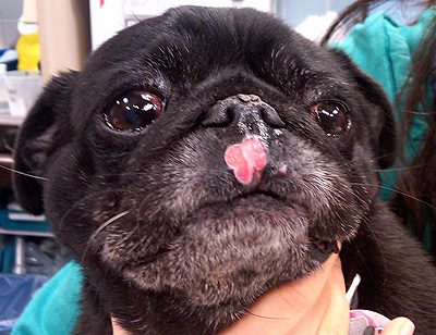
- Some breeds have a higher incidence, for example brachiocephalic breeds (those with a short, wide head and a short nose), such as boxers, bulldogs and pugs
- It is also prevalent in Boston Terriers, Beagles, Labrador Retrievers, and Golden Retrievers
- Twenty to thirty percent of mast cell tumours have a mutation in a specific gene called c-kit
Environmental factors may predispose their development, including:
- Chronic skin inflammation
- Irritants on the skin, applied chronically
Age:
- Onset can occur at any age, but the likelihood increases with increasing age
- The average age of affected dogs is 8 to 9 years
How are mast cell tumours diagnosed?
Often the first indication of a mast cell tumour is the discovery of a lump on or under the dog’s skin. As any lump may be abnormal and even cancerous, you should immediately take your dog to the vet for examination. Where there are no lumps or obvious symptoms, mast cell dog tumours may be discovered incidentally during a routine veterinary examination.
Diagnostic tests may include:
- Preliminary biopsy – Because mast cell tumours in dogs can appear in many shapes, sizes and locations and many of the symptoms can be associated with other conditions, a biopsy (or fine needle aspiration) is usually performed so that the tissue sample can be examined through a microscope.
- Surgical tissue biopsy – after it has been removed, the mast cell dog tumour is examined for classification and grading of the cells and identifying the stage of the disease. This is necessary for determining treatment and prognosis.
- A sample obtained from draining the nearest lymph node, or bone marrow, kidney or spleen may be examined
- X-ray and / or ultrasound imaging of the chest and abdomen may assist in identifying the stage and location
- Complete blood count
- Blood chemistry panel
- Urinalysis (urine sampling)
- If other organs are swollen or show signs of being affected, assessment by ultrasound, aspiration and/or biopsy
Grading of mast cell tumours / mastocytoma:
- Grading of tumours identifies the degree of malignancy
- Determined by biopsy (examination of a portion of the affected tissue by a pathologist)
- Mast cell tumours have three grades:
- Grade 1 – occur on in the skin, considered benign, tend not to spread
- Grade 2 – extend into the tissue below the skin, have potential for local metastasis or spread
- Grade 3 – extend into areas deep below the skin, have a high potential for metastasis / spread, very aggressive
Staging of mast cell tumours / mastocytoma:
- Staging of tumours identifies how they have spread in the body
- Determined by examination of the tumour and its closest lymph nodes
- Staging is based on the number of tumours, lymph nodes affected, and how much it has metastasised
Stage 1 – a single tumour without metastasis
- A good to excellent prognosis with appropriate treatment
Stage 2 – a single tumour with metastasis into the surrounding lymph nodes
- a very good prognosis with appropriate treatment
Stage 3 – multiple skin tumours, or a large tumour that has invaded subcutaneously, with or without lymph node involvement
- because of the presence of malignancy and metastasis, a guarded to grave prognosis
Stage 4 – one or more tumours, with metastasis to the skin or an organ or wide spread mast cell presence in the blood and lymph node involvement
- because of the presence of malignancy and metastasis, a guarded to grave prognosis
About 50% of mast cell tumours in dogs are malignant, meaning that if left untreated they will usually spread to other parts of the body, such as the spleen, liver, lymph nodes and bone marrow, and result ultimately in death.
The prognosis for dogs with a mast cell tumour depends on the grade and stage of the tumour at the time of diagnosis, with those staged/graded 1 to 2 usually having a high rate of success. However, tumours of higher grade and stage, tumours that have spread to other parts of the body and those that have formed in areas other than the skin, generally have a much poorer prognosis, depending on how well they respond to chemotherapy and radiation treatments.
More than 15% of dogs diagnosed with a mast cell tumour will develop further mast cell tumours in their lifetime. Therefore it is essential to undertake regular inspections of your dog’s skin for any signs of new tumours for the rest of its life.
Treatment for mast cell tumours
Each case of mast cell tumour is individually evaluated and treated, depending on the diagnosed grade (the degree of malignancy) and stage (the likelihood of spread). The vet’s treatment preferences will also play a role. The prognosis depends on the grade and stage of the disease at the time of diagnosis, as well as the dog’s response to the selected treatments.
Depending on the size and location of the dog tumour, treatment usually requires surgical removal, which may be followed by chemotherapy, radiation and / or steroids.
The goals of treatment are:
- To remove the tumour and any other affected areas
- To prevent metastasis
- To improve, maintain or at least manage the affected dog’s quality of life
Surgical removal
The primary treatment for dogs with mast cell tumours is surgical removal.
- Treatment for Grade I and Grade II tumours:
- Surgery is the treatment of choice, and if performed correctly, is highly successful for curing low grade tumours
- It is important that the tumour is carefully removed and a large area of ‘healthy’ tissue around the tumour is also removed. It is often difficult to determine exactly where the tumour begins and healthy tissue starts, so a wide margin (a large portion of healthy tissue surrounding the tumour) is removed to ensure that all cancerous tissue at the site is eliminated.
- Nearby lymph nodes are also generally removed, because they are the first place where the mast cell tumour spreads.
- Further treatment may not be required.
- Treatment for more complex tumours, larger tumours and where it is not possible to remove the tumour with good margins:
- Surgery is performed to remove as much of the tumour and margins as possible, followed by radiation, chemotherapy and / or immunotherapy and in some cases, further surgery to remove more tissue.
- Grade 3 tumours are invariably malignant and after surgical removal there is a high chance of regrowth and of spreading to lymph nodes, internal organs and even bone marrow. Aggressive surgical removal of the tumour and affected lymph nodes and any other affected organs is required, followed by other treatments.
- If there is a generalised spread or metastasis of the tumour cells to other parts of the body, surgery may have minimal benefit.
Radiation therapy
Radiation therapy can clear up any remaining tumour cells after surgery, and is a likely treatment option in the following situations:
- If surgical removal of the mast cell tumour is not possible
- If not enough of the surgical margin can be removed
- After surgery where it may reduce the incidence of reoccurrence and / or increase survival rates
- Where tumours have not spread to multiple areas of the body
- Where the tumour is on an extremity (tumours on an extremity respond better to radiation than tumours on the trunk)
Chemotherapy
Chemotherapy following surgery is recommended in certain cases, such as:
- After surgery, for the prevention or reduction of the chance of further metastasis
- If surgery is not performed because cancer cells have already spread considerably to other parts of the body, chemotherapy can offer short-term benefit (up to two months)
- If surgery is performed but the primary tumour cannot be entirely removed, chemotherapy can offer a short-term respite from the effects of the disease (one to four months)
Immunosuppressive steroid administration / Anti-histamines and other medications for the systemic effects
- Mast cell tumours release histamines into the blood stream, therefore antihistamines (e.g. Benadryl) may be prescribed to alleviate symptoms (excessive histamines in the body can have a dramatic effect on the organs)
- Acid blockers (e.g. Pepcid, Prilosec) counteract the acid increasing substance produced by mast cell tumours
- Steroids (e.g. Prednisone) can kill cancerous mast cells, reduce inflammation and mitigate the effects of the chemicals released by mast cell tumours, however these drugs can have unpleasant side effects, such as nausea, vomiting, diarrhea
- A combination of these drugs may be prescribed where multimodality therapy is indicated, usually where there are high grade mast cell tumours or metastasis to nearby lymph nodes or beyond.
Supportive and ongoing care
Dogs that have had mast cell tumours have a greater risk for developing additional tumours, therefore the following courses of action are likely on an ongoing basis:
- Vigilant inspection of the dog’s skin for any signs of new masses (for the rest of the animal’s life)
- Microscopic analysis of any new lumps
- Regular check-ups by the vet
- Providing a healthy, immune boosting diet
Overview
Mast cells are found in large quantities in a dog’s skin. Mast cell tumours, the most common type of dog skin cancers, often appear as isolated lumps on the skin, but can present in many different locations and vary widely in appearance and growth patterns. While many are benign, around 50% are cancerous and can spread to other locations, which can be fatal.
It is difficult to detect mast cell tumours visually, therefore it is essential to have any lumps or bumps examined by the vet and diagnostically tested. If found to be a mast cell tumour, treatment for this dog skin cancer is likely to consist of surgical removal, radiation and / or chemotherapy and / or systemic therapies. Treating mast cell tumours when they are small and localised has a high rate of success. However, tumours of higher grade and stage generally have a much poorer prognosis, depending largely on the nature and extent of the metastasis and how they respond to chemotherapy and radiation treatments.
Bow Wow Meow Pet Insurance can help protect you and your dog should an unexpected trip to the vet occur.
-
Find out more about our dog insurance options
-
Get an online pet insurance quote
Bow Wow Meow is proud to have been awarded winner of Canstar’s ‘Most Satisfied Customers’ Award in the Pet Insurance category for both 2024 and 2025!
Bow Wow Meow is proud to be the only Pet Insurer that has been awarded Product Review’s Top Rated Pet Insurance every year from 2018 to 2026! This is based on 3,193 independent customer reviews (as at 09/04/2026), with an overall rating of 4.5 stars.

We also have the following ratings on other review platforms:
Google Review rating of 4.5 stars (based on 1,358 reviews)
Trust Pilot rating of 4.6 stars (based on 555 reviews)
Bow Wow Meow has been chosen as Winner of 4 categories in the 2026 Finder Pet Insurance Customer Satisfaction Awards!


– Most Trusted Pet Insurance
– Easiest to Claim Pet Insurance
– Most Recommended Pet Insurance
– Legendary Service Pet Insurance
We also received Highly Commended Awards for Loved Brand Pet Insurance and Best Value Pet Insurance.
Bow Wow Meow is an Australian-owned and operated pet insurance provider. Our focus is purely on pets, and we are proud to have helped provide peace of mind to over 200,000 Aussie pet owners since 2008.
More information
https://www.petwave.com/Dogs/Health/Mast-Cell-Tumors.aspx
https://blogs.webmd.com/pet-tales/2011/01/twenty-five-years-of-mast-cell-tumors.html
https://pets.webmd.com/dogs/dogs-and-skin-cancer#1
https://www.petmd.com/blogs/thedailyvet/jintile/2013/june/the-dreaded-mast-cell-tumor-30465
https://www.petmd.com/blogs/fullyvetted/2012/aug/mast_cell_tumors_in_dogs-26719
https://www.petmd.com/dog/conditions/cancer/c_dg_mast_cell_tumor
https://www.petcoach.co/article/mast-cell-tumors/
https://www.ncbi.nlm.nih.gov/pubmed/21961635
https://www.vet.upenn.edu/docs/default-source/ryan/oncology-handouts/final-canine-mct.pdf?sfvrsn=4