Mast cell tumours in dogs: symptoms, diagnosis and treatment



Summary
Mast cell tumours (MCTs) are among the most common skin tumours diagnosed in dogs. They usually appear as a lump on or beneath the skin, but their appearance can vary enormously, from a small, seemingly harmless bump to a red, swollen or ulcerated mass. It is not possible to identify a mast cell tumour simply by looking at it.
The behaviour of mast cell tumours also varies considerably. Many low-grade, localised tumours can be successfully treated with surgery, while high-grade tumours are more likely to recur or spread to other parts of the body. Any new, changing or unexplained lump on your dog should therefore be checked by a veterinarian.
What is a mast cell tumour?
Mast cells are immune cells (a type of white blood cell) that are found throughout the body, particularly the skin. They play an important role in inflammation and allergic reactions by releasing substances including histamine.
Mast cell tumours in dogs, also called MCTs or mastocytomas, are one of the most common type of dog skin cancers. A mast cell tumour develops when mast cells begin multiplying abnormally and form a mass. In dogs, most mast cell tumours arise in the skin or tissue immediately beneath it, although mast cell disease can occasionally involve internal organs.
The tumours often appear small and insignificant, but in severe cases they can be very serious and even life threatening. Mast cells can also release large quantities of histamine and other inflammatory substances. This can cause redness and swelling around the tumour and, in some dogs, gastrointestinal problems such as stomach or intestinal ulceration.
Expert advice from Dr Felicia:
Mast cell tumours are frequently encountered in vet practices, particularly in brachycephalic dogs. Thankfully most of them are low grade and surgery with clean margins is curative, though some dogs are prone to developing developing more mast cell tumours.
What does a mast cell tumour look like on a dog?
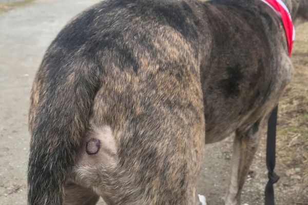
Mast cell tumours are sometimes called “the great pretenders” because they can resemble many other skin lumps and lesions.
A mast cell tumour may be:
- a small lump within or beneath otherwise normal-looking skin
- soft or firm to touch
- smooth, raised or irregular
- hairless or covered with normal fur
- red, inflamed or itchy
- ulcerated or weeping
- slow-growing or rapidly enlarging
- a lump that seems to change size from one day to the next.
Some MCTs become temporarily larger, redder or more irritated when they are touched or disturbed because mast cells release histamine and other substances into the surrounding tissue.
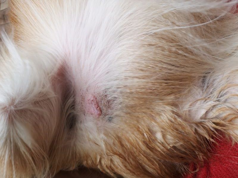
A mast cell tumour cannot be reliably distinguished from a harmless lump by appearance or touch alone. A small or apparently insignificant lump can still be an MCT, which is why veterinary assessment and sampling are important.
Symptoms of mast cell tumours
Mast cell tumours in dogs are extremely variable in their location, appearance, and growth pattern. Many affected dogs show no signs of illness or discomfort – the only sign is a lump on or beneath the skin, and they otherwise appear completely well.
Depending on the tumour and the substances released by its mast cells, other signs may include:
- redness, swelling or itching around the lump
- sudden enlargement or fluctuation in the size of the lump
- ulceration or bleeding from the tumour
- vomiting
- reduced appetite
- diarrhoea
- abdominal discomfort
- dark, tarry stools caused by gastrointestinal bleeding
- enlarged lymph nodes
- weakness or lethargy.
Severe mast-cell degranulation can occasionally result in a widespread inflammatory or allergic-type reaction, including collapse or shock.
Expert advice from Dr Felicia:
Being able to examine the cells comprising the mass is crucial for identifying mast cell tumours. While this is usually straightforward for many dogs, some locations can be hard to sample in a conscious animal (eg. face, feet) and a sedation may be required to be able to safely aspirate the lump.
When should you see the vet?
Any new or changing lump should be assessed by a veterinarian, particularly if it is growing, changing size, becoming red or ulcerated, or bothering your dog.
Seek prompt veterinary care if your dog has a lump together with repeated vomiting, marked weakness, black or tarry stools, collapse, breathing difficulty or other signs of severe illness.

What causes mast cell tumours in dogs?
The exact cause of mast cell tumours is not known. As with many cancers, their development is likely to involve a combination of genetic and biological factors rather than one single cause.
MCTs can occur at any age but are seen more commonly in middle-aged and older dogs. Certain breeds appear to have an increased risk, including Boxers, Pugs, Boston Terriers, Labradors and Golden Retrievers, although any dog can develop a mast cell tumour.
Breed alone does not determine how an individual tumour will behave.
How are mast cell tumours diagnosed?
Often the first indication of a mast cell tumour is the discovery of a lump on or under the dog’s skin. As any lump may be abnormal and even cancerous, you should immediately take your dog to the vet for examination. Where there are no lumps or obvious symptoms, mast cell dog tumours may be discovered incidentally during a routine veterinary examination.
Fine-needle aspiration
A veterinarian can often diagnose an MCT using a fine-needle aspirate (FNA). A small needle is inserted into the lump to collect cells, which are then examined under a microscope.
Because mast cell tumours in dogs can appear in many shapes, sizes and locations and many of the symptoms can be associated with other conditions, a fine needle aspirate is usually performed so that the tissue sample can be examined through a microscope. Mast cells have distinctive granules that frequently make MCTs relatively straightforward to recognise on cytology.
Histopathology
If the tumour is surgically removed or biopsied, the tissue is sent to a veterinary pathologist for histopathology. This is necessary for determining treatment and prognosis.
Histopathology provides important information including:
- confirmation of the diagnosis
- tumour grade
- features associated with biological behaviour
- whether tumour cells extend to the surgical margins.
Mast cell tumour grade and stage: what is the difference?
Grade and stage describe different aspects of the tumour.
Tumour grade
Grade describes how the tumour cells look and behave under the microscope and helps predict how aggressively the tumour is likely to behave.
The Kiupel grading system classifies cutaneous mast cell tumours as either:
- Low grade – generally associated with a lower risk of aggressive behaviour and a more favourable prognosis.
- High grade – more likely to recur, spread and behave aggressively.
Expert advice from Dr Felicia:
Thankfully the majority of cutaneous MCTs are categorised as Grade 1, with excision considered curative. The other grades require some further discussion about next steps and further therapy.
Tumour stage
Stage describes how far the cancer has spread.
Depending on the characteristics of the tumour and the dog’s clinical findings, staging may include examination or sampling of the lymph nodes that drain the tumour, blood tests and imaging such as abdominal ultrasound.
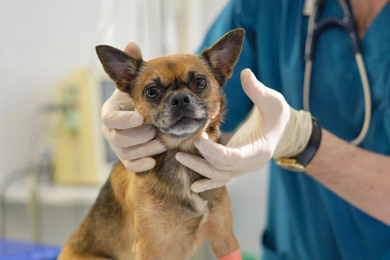
Lymph-node assessment is particularly important because the first lymph node draining the tumour – known as the sentinel lymph node – may contain tumour cells even when it is not enlarged.
Not every dog requires exactly the same staging tests. Your veterinarian or veterinary oncologist will recommend testing according to the tumour’s grade, location, clinical appearance and other findings.
Treatment for mast cell tumours in dogs
Treatment depends on factors including the tumour’s size and location, its grade, whether it has been completely removed and whether there is evidence that it has spread.
Surgery
Surgical removal is the main treatment for many localised mast cell tumours.
Because microscopic mast cells may extend beyond the visible edge of the lump, the surgeon will aim to remove an appropriate margin of normal-looking tissue around and beneath the tumour where possible.
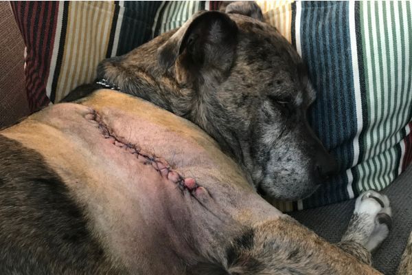
The removed tissue is then examined by a pathologist to determine the tumour grade and assess the surgical margins.
For many low-grade MCTs that are completely removed, surgery may be the only treatment required.
Expert advice from Dr Felicia:
Tumours on the face and feet are difficult to remove with ideal margins, and are considered more complex on this basis.
Further surgery or radiation therapy
If tumour cells extend to or close to the surgical margin, the next step depends on the tumour’s biological risk, location and other clinical factors.
Options may include:
- further surgery to obtain wider margins
- radiation therapy to improve local tumour control
- careful monitoring in selected lower-risk cases.
These decisions are best made with your veterinarian or veterinary oncologist rather than assuming that every incompletely removed tumour requires the same treatment.
Chemotherapy
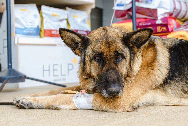
Chemotherapy may be recommended for some high-grade mast cell tumours, tumours that have spread to lymph nodes or other organs, or cases considered to have a significant risk of metastasis.
Intra-tumoral injection
In patients where surgery is not practical due to location or anaesthesia concerns, an intra-tumoral injection can destroy tumour tissue, causing the body to initiate an inflammatory response to the region.
Disadvantages of intra-tumoral injection include significant local pain and inflammation to the site during the treatment, as well as no histopathology to determine the tumour grade.
Medications to manage mast-cell effects
Because mast cell tumours can release histamine and other inflammatory substances, veterinarians may prescribe medications such as antihistamines, acid-suppressing drugs or corticosteroids in selected cases.
These medications should only be given under veterinary direction.
Expert advice from Dr Felicia:
Deciding whether to proceed with full chemotherapy or palliation may depend any other health concerns your dog may have or their ability to tolerate these medications, and your vet will be able to advise on what is best for your pet.
Supportive and ongoing care
Dogs that have had mast cell tumours have a greater risk for developing additional tumours, therefore the following courses of action are likely on an ongoing basis:
- Vigilant inspection of the dog’s skin for any signs of new masses (for the rest of the animal’s life)
- Microscopic analysis of any new lumps
- Regular check-ups by the vet
What is the prognosis for a dog with a mast cell tumour?
There is no single life expectancy for a dog diagnosed with an MCT. The prognosis depends on factors including:
- whether the tumour is low or high grade
- whether it can be completely treated locally
- tumour size and location
- lymph-node involvement
- whether the cancer has spread elsewhere
- microscopic features such as mitotic activity
- the dog’s response to treatment.
Many dogs with low-grade, localised mast cell tumours that receive appropriate local treatment have an excellent prognosis, and treatment may be curative.
High-grade tumours and MCTs that have spread to lymph nodes or internal organs have a more guarded prognosis and usually require more intensive treatment and monitoring.

Can mast cell tumours come back?
A mast cell tumour can recur at the original site if tumour cells remain, particularly with biologically aggressive tumours.
Dogs that have developed one MCT can also develop new, unrelated mast cell tumours elsewhere on their body. A new lump does not automatically mean that the original cancer has spread.
After treatment, your veterinarian may recommend regular physical examinations and ongoing checks of your dog’s skin so that any new or changing lumps can be assessed promptly.
Can mast cell tumours in dogs be prevented?
There is currently no proven way to prevent most mast cell tumours.
The most useful action owners can take is to become familiar with their dog’s skin and body and have new or changing lumps assessed early.
Do not rely on appearance alone or wait for a lump to become large before having it checked.
In summary
Mast cell tumours are common canine skin tumours with extremely variable behaviour. They may look like an ordinary skin lump, and it is usually impossible to determine how serious a mass is simply by looking at it.
Fine-needle aspiration can often identify an MCT, while histopathology provides essential information about grade and surgical margins. Many low-grade, localised tumours can be treated very successfully, particularly when identified early, while high-grade or metastatic tumours require more extensive treatment.
If you discover a new lump or notice a change in an existing one, arrange a veterinary examination rather than waiting to see what it does.
Bow Wow Meow Pet Insurance can help protect you and your dog should an unexpected trip to the vet occur.
-
Find out more about our dog insurance options
-
Get an online pet insurance quote
Bow Wow Meow is proud to have been awarded winner of Canstar’s ‘Most Satisfied Customers’ Award in the Pet Insurance category for both 2024 and 2025!
Bow Wow Meow is proud to be the only Pet Insurer that has been awarded Product Review’s Top Rated Pet Insurance every year from 2018 to 2026! This is based on 3,193 independent customer reviews (as at 09/04/2026), with an overall rating of 4.5 stars.

We also have the following ratings on other review platforms:
Google Review rating of 4.5 stars (based on 1,358 reviews)
Trust Pilot rating of 4.6 stars (based on 555 reviews)
Bow Wow Meow has been chosen as Winner of 4 categories in the 2026 Finder Pet Insurance Customer Satisfaction Awards!




– Most Trusted Pet Insurance
– Easiest to Claim Pet Insurance
– Most Recommended Pet Insurance
– Legendary Service Pet Insurance
We also received Highly Commended Awards for Loved Brand Pet Insurance and Best Value Pet Insurance.
Bow Wow Meow is an Australian-owned and operated pet insurance provider. Our focus is purely on pets, and we are proud to have helped provide peace of mind to over 200,000 Aussie pet owners since 2008.
Sources
“Mast Cell Tumors”, American College of Veterinary Surgeons. https://www.acvs.org/small-animal/mast-cell-tumors/. Accessed 15 September 2026.
“Mast Cell Tumors in Dogs”, VCA Animal Hospitals. https://vcahospitals.com/tanglewood/know-your-pet/mast-cell-tumors-in-dogs. Accessed 15 September 2026.
“Lymphocytic, Histiocytic, and Related Cutaneous Tumors in Animals”, Merck Veterinary Manual. https://www.merckvetmanual.com/integumentary-system/tumors-of-the-skin-and-soft-tissues/lymphocytic-histiocytic-and-related-cutaneous-tumors-in-animals. Accessed 15 September 2026.
Willmann M, et al. “Proposed Diagnostic Criteria and Classification of Canine Mast Cell Neoplasms: A Consensus Proposal”, Frontiers in Veterinary Science, 2021. https://pmc.ncbi.nlm.nih.gov/articles/PMC8702826/. Accessed 15 September 2026.
Blackwood L, et al. “Canine cutaneous and subcutaneous mast cell tumours: a narrative review”, Journal of Small Animal Practice, 2022. https://pubmed.ncbi.nlm.nih.gov/34671978/. Accessed 15 September 2026.
Kiupel M, et al. “Value, Limitations, and Recommendations for Grading of Canine Cutaneous Mast Cell Tumors: A Consensus of the Oncology-Pathology Working Group”, Veterinary Pathology, 2021. https://pubmed.ncbi.nlm.nih.gov/33888024/. Accessed 15 September 2026.
“Targeted Antineoplastic Agents in Animals”, Merck Veterinary Manual. https://www.merckvetmanual.com/pharmacology/antineoplastic-agents/targeted-antineoplastic-agents-in-animals. Accessed 15 September 2026.
“STELFONTA – tigilanol tiglate”, Australian Pesticides and Veterinary Medicines Authority, 2021. https://www.apvma.gov.au/sites/default/files/gazette_20210309.pdf. Accessed 15 September 2026.