
Alimentary foreign body in dogs and cats

What is an alimentary foreign body in dogs and cats?
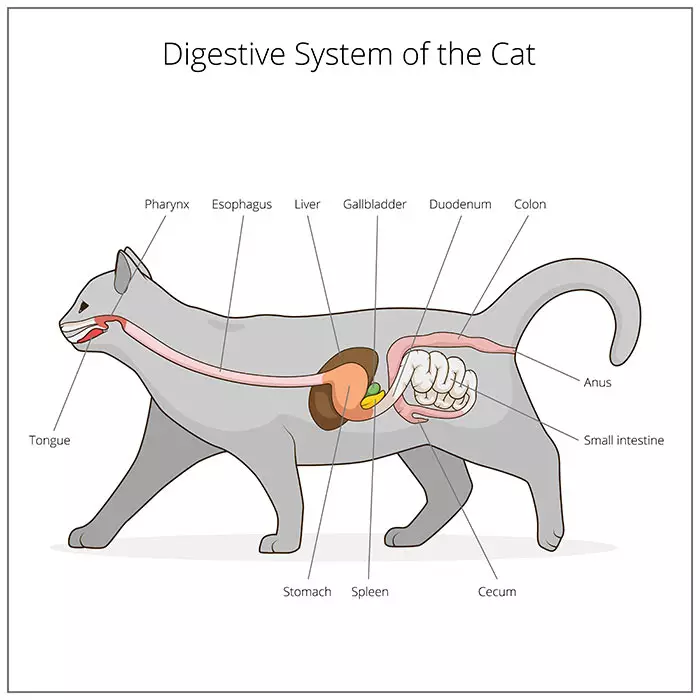
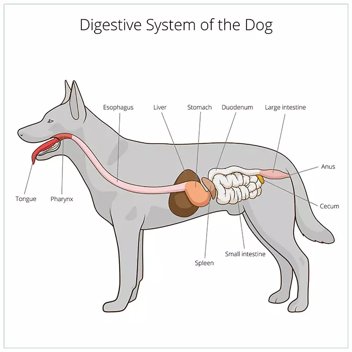
The alimentary canal, also known as the digestive or gastrointestinal tract, is the long tube of the digestive system that extends from the mouth to the anus, through which food passes, digestion takes place and wastes are eliminated. The oesophagus is in the upper section of the alimentary canal, extending from the throat to the stomach, while the bowel, or intestines, is the part of the alimentary canal below the stomach.
The term ‘foreign body’ refers to any non-food object located within the alimentary canal. Pets have the tendency to play with, or chew on, non-food items and these objects can be inadvertently swallowed. While smaller foreign bodies may pass through the gastrointestinal system without becoming stuck or causing damage, larger objects can ‘get stuck’ and cause an obstruction or blockage in any part of the alimentary canal. Most commonly, obstruction occurs in the small intestine because of its narrow diameter. This condition is also known as a bowel obstruction in dogs and cats.
Foreign bodies lodged in the intestines or stomach and can have life-threatening consequences. Dogs and cats of any age can suffer from foreign body obstructions but young animals under two years of age are more frequently afflicted because of their naturally curious natures.
Symptoms of alimentary foreign bodies in dogs and cats
Clinical signs can vary significantly depending on the degree of obstruction, the location within the alimentary canal, the duration of the obstruction, and the type of foreign body ingested.
Symptoms of obstruction of the oesophagus in dogs and cats may include:
- Regurgitation of food and water
- Cervical discomfort
- Respiratory distress
- Retching
- Ptyalism (excessive production of saliva)
- Anorexia (decrease or loss of appetite)
- Restlessness
Symptoms of bowel obstruction in dogs and cats are largely similar, and include:
- Vomiting
- Decreased appetite
- Lethargy
- Abdominal pain
- Hematemesis (vomiting of blood)
- Anorexia (decrease or loss of appetite)
- Dehydration
- Constipation
- Straining to produce a bowel movement
- Blood in the faeces
- Reduced volume of faeces
- Diarrhoea
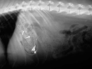
Abdominal radiograph (X-ray) of a dog that ate some nails which you can see in his stomach.
Source: https://www.medvetforpets.com/gastrointestinal-foreign-bodies-fb-dogs-cats/
Causes of an alimentary foreign body in dogs and cats
Dogs and cats tend to eat things that they should not, and while smaller foreign bodies can pass through the body in the stool, others will become lodged somewhere along the alimentary canal.
Dogs, especially young dogs, commonly chew on objects, so if you notice the recent disappearance of an object, keep in mind the possibility of a foreign object in the dog’s stomach or other location within the alimentary canal.
Common foreign objects found in dogs’ stomachs include:
- Bones
- Stones
- Plastics, food wrappings
- Rawhide
- Toys and balls
- Dental chews (greenies)
- Fish hooks
- Coins
- Towels
- Socks, underwear and nylons
- Rubber objects

Common foreign objects found in cats’ stomachs include:
- Needles
- String
- Toys
- Elastics
- Plastic
- Hairballs
What are linear foreign bodies?
An especially dangerous type of foreign body, most commonly found in cats, is referred to as a linear foreign body, which is a long, thin object such as string, yarn, or tinsel. One end of the linear object can become lodged, for example at the base of the tongue, while the other end trails down the remainder of the gastrointestinal tract, unable to be eliminated. This can cause a crawling or bunching of the intestines, which can lead to perforation of the alimentary canal and spillage of intestinal contents into the abdomen, with life-threatening complications. Sometimes linear foreign bodies can become lodged in multiple locations in the alimentary tract if they are composed of string, fabric or carpet.
Problems caused by the ingestion of a foreign body vary, depending on:
- The features of the foreign body:
- Items made of a metal such as lead or zinc, including older coins or building materials, can cause anaemia and systemic toxicity
- Round smooth objects, such as balls, may cause complete intestinal obstruction and pressure damage to the intestinal wall
- Sharp objects, such as bones or needles, may penetrate the intestinal wall and cause septic peritonitis
- The degree of obstruction that is caused:
- There may be a partial obstruction or complete obstruction
- The duration that the foreign body has been present:
- The longer the object remains in the body, the greater the chances of serious complications and even death
- The location of the foreign body:
- Oesophageal foreign bodies often cause regurgitation, cervical discomfort, and in some cases, respiratory distress
- If the stomach or intestines are obstructed, severe dehydration and electrolyte changes can occur
- If the foreign body moves into the intestines:
- It may cause damage to the intestinal tract itself through compression or obstruction, such as perforation of the intestinal wall with subsequent peritonitis and sepsis, which carry a guarded prognosis and require additional operative and postoperative treatment
- It can cause intestinal oedema (a condition characterized by an excess of watery fluid collecting in the cavities or tissues of the body)
How is an alimentary foreign body in dogs and cats diagnosed?
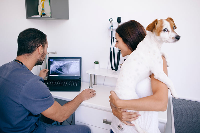
Foreign body ingestion should always be investigated in a dog or cat presenting with symptoms such as a lack of appetite, acute vomiting, chronic diarrhoea and weight loss, regardless of age and history. Diagnosis is usually made via physical examination, radiography and / or ultrasonography. Your vet may also perform blood tests, including a complete blood count (CBC), serum chemistry and urinalysis, to help rule out other possible causes.
Physical examination
Abdominal palpation is important in the diagnosis of an obstruction, but additional diagnostics are usually required for confirmation. Palpation of the animal’s abdomen or throat can sometimes reveal the presence of the foreign body itself, or pain in the region of the obstruction.
Where a foreign body is lodged in the oesophagus, physical examination of the throat may reveal ptyalism (an increased amount of saliva in the mouth) and discomfort in the cervical spine (neck).
Where there is a gastrointestinal foreign body, abdominal palpation may not reveal anything, or palpation may elicit abdominal pain. Gas build-up in front of the obstruction with distension of the intestines may be detected.
Radiography
Abdominal, and occasionally thoracic, radiographs (X-rays) are regularly performed in both dogs and cats in the diagnosis of alimentary foreign bodies. Radiographs can be used to identify the site of the obstruction and are especially helpful if the foreign body contains bone, metal or rock which show up characteristically on X-ray.
In dogs, abdominal radiographs are the most common diagnostic test performed to find evidence of a foreign object in a dog’s stomach. Often the foreign body cannot be seen on the x-ray, but the consequences of the foreign body obstruction are visible. These include fluid and gas building up behind or within the foreign body.
In cats, linear foreign bodies are most commonly diagnosed using X-rays. Although the object itself may not be visible on X-rays, linear foreign bodies can cause the intestines to bunch in a way that may be observed on X-rays, in what is known as a “string-of-pearls” appearance to the intestines, or there may be observable abnormal gas patterns within the intestines.
Positive contrast radiographs
Foreign objects in a dog’s or cat’s stomach that made of organic material, such as corn cobs, string and hair balls, may be the same density as soft tissue on x-rays and are frequently not identifiable on routine X-rays. In such cases, contrast radiography using barium, a dye-like material, to highlight the inside of the stomach and intestines can be helpful in outlining the obstruction. However, while barium administration can define some bowel obstructions in dogs and cats, it does not usually provide a definitive diagnosis. Note that the ingestion of contract dye is not recommended if an oesophageal obstruction is suspected, as there is a relatively high risk of aspiration or choking.
Ultrasound
If radiography results are inclusive, an abdominal ultrasound may be performed to aid in identifying a bowel obstruction in dogs and cats. Abdominal ultrasonography is considered the most useful non-invasive method for diagnosis of small intestinal foreign bodies and can strongly suggest the presence of a bowel obstruction. Because ultrasound gives a 3D view of the intestines, it may be more effective than X-rays when looking for a foreign object in a dog’s stomach or a linear foreign body in a cat’s intestines, for example. However, ultrasonography is rarely useful in cases of oesophageal foreign bodies.
Using ultrasound, a shadowing object may be observable within the small intestine, typically causing some degree of dilation of the intestine at the site of obstruction. However, gas and food material can create shadowing that masks foreign objects. Abdominal ultrasound is also useful to rule out perforation of the colon, which can lead to septic peritonitis.
Bowel obstruction in dogs and cats can be challenging to definitively diagnose on both radiographs and ultrasound examination, as there is often gas and food material present in the stomach that can cause shadowing and masking of foreign objects in the stomach. However, both radiographs and ultrasound can strongly suggest the presence of a gastric foreign body. If food and/or gas are present, then imaging can be repeated after a period of fasting, however there is a risk that this could allow a possible foreign object to enter the small intestine.
Prognosis
Most uncomplicated gastrointestinal foreign bodies have an excellent prognosis; however, delayed presentation, diagnosis and surgical intervention will worsen the prognosis considerably. If not removed rapidly, bowel obstructions in dogs and cats are associated with high morbidity and mortality.
The location of the foreign body also may indicate a poorer outcome. Foreign objects in dogs’ stomachs, as well as cats’, can enter the small intestine and the resultant obstruction can lead to perforation of the intestinal wall, with subsequent peritonitis and sepsis, which carry a guarded prognosis and require additional operative and postoperative treatment.
Additionally, intestinal obstruction, protracted vomiting, and diarrhoea can cause significant metabolic changes within the body that need to be addressed post removal. Furthermore, many foreign bodies are made of materials that are potentially toxic when absorbed, for example, lead and zinc may lead to profound systemic disease if a sufficient quantity is ingested.
Treatment for alimentary foreign body in dogs and cats
Oesophageal foreign bodies are considered an emergency and require immediate removal in all cases. Most foreign bodies located in the upper alimentary canal may be retrieved with the use of an endoscope; however, if unsuccessful, surgical exploration and removal is required.
Foreign bodies in the lower alimentary tract are not always an emergency, and diagnostics may indicate that conservative management is an appropriate initial response. In other words, surgical intervention is not always required for a cat’s or dog’s intestinal blockage. Some items are small and smooth enough to pass through the gastrointestinal tract without causing damage or becoming lodged. The decision on whether to remove the foreign body depends on location, clinical signs, time since ingestion of the item, and size, shape and nature of the foreign body.
Conservative management
- The foreign body is not removed through intervention and is allowed to pass through the body and evacuated in the faeces
- Generally more suited to asymptomatic animals that have ingested small, smooth foreign bodies
- Some small, sharp gastrointestinal foreign bodies, such as pins, sewing needles and fish hooks, that are found in asymptomatic animals may be treated conservatively and will pass without issue through the alimentary canal
- The pros and cons of removal of smaller objects should be discussed in all cases, so that owners can make educated decisions about whether to follow a conservative approach or pursue removal.
Endoscopy
- Endoscopy is a medical procedure that allows your vet to inspect and observe the inside of the body without performing major surgery.
- Smaller foreign bodies that become lodged in the upper gastrointestinal tract (mouth, oesophagus, and stomach) and may be removed with the use of a flexible endoscope.
- An endoscope is a long, usually flexible tube with a lens at one end and a video camera at the other
- There are a variety of endoscopic grasping forceps, nets and snares that can be used to remove the object
- The majority of oesophageal and gastric foreign bodies can be removed endoscopically
- However, if the foreign body has a very sharp surface (such as a razor blade) or sharp point (such as a fish hook), endoscopic removal may pose too high a risk to removal through the oesophagus
Surgery
- Foreign body obstructions are among the most common causes for emergency veterinary surgery
- Laparotomy, a surgical incision into the abdominal cavity, may be performed for diagnosis and surgical exploration of the pet’s abdomen
- If the foreign body has lodged within the stomach or intestines, a gastrotomy (opening the stomach) or enterotomy (opening the intestine) may be performed
- Gastrotomy, or cutting open the stomach, is a relatively simple procedure to remove foreign material from the stomach, and most gastric foreign bodies can be removed rapidly and without complication
- Enterotomy, or cutting open the intestine, is a relatively quick procedure which is indicated when a small intestinal foreign body has been diagnosed. However, many cases will present with multiple foreign bodies, which require several enterotomies.
- Colonoscopy remains the most effective and least invasive method of removal for colonic foreign bodies.
- Occasionally, foreign bodies will become lodged in the oesophagus at the base of the heart or at the diaphragm, which may require thoracic (chest) surgery to remove them
- Immediate removal is indicated for large objects, objects with sharp points or sharp surfaces, irregular objects, and caustic containing material such as batteries or pennies
- If there are many linear foreign bodies and completely obstructed intestines, there may be severe intestinal damage so that multiple enterotomies are required
- If a section of bowel is irreversibly damaged, an intestinal resection and anastomosis (procedure to remove a segment of the intestines and reattach the healthy ends) may be performed
- In cases of chronic obstruction or the presence of a large or sharp foreign body, necrosis and laceration of the intestines or stomach around the foreign body can result in leakage of intestinal contents into the abdomen, causing infection (peritonitis) which puts the animal at high risk of septicaemia and death
- Once the foreign body has been surgically removed, the animal is maintained on fluids and may require hospitalisation for several days while food and water are re-introduced, electrolytes are normalised and bowel movements are monitored
Overview
Bowel obstruction in dogs and cats, as with obstruction within other locations of the alimentary tract, occurs when the animal eats a non-food foreign object and it cannot pass through. Obstruction in dogs and cats can be caused by ingestion of common objects such as toys, socks, string, bones and balls. Symptoms of bowel obstruction in dogs and cats can include vomiting, anorexia (refusal to eat), dehydration, and lethargy; or the animal may be asymptomatic.
Foreign body obstructions are among the most common causes for emergency veterinary surgery in dogs. X-rays and abdominal ultrasound can be very helpful in identifying a foreign object in a dog’s stomach or intestines. If not removed rapidly, a bowel obstruction in dogs can cause damage to a section of the alimentary tract and have a poor prognosis.
Although not always practically possible, preventing access to hazards will greatly reduce the risk of your cat or dog developing this life-threatening condition. Prompt intervention in cases of gastrointestinal foreign body obstruction in dogs and cats is essential to give your pet the best chance of survival.
Bow Wow Meow Pet Insurance can help protect you and your pet should an unexpected trip to your vet occur.
- Find out more about our dog insurance options
- Find out more about our cat insurance options
- Get an instant online pet insurance quote
Bow Wow Meow is proud to have been awarded winner of Canstar’s ‘Most Satisfied Customers’ Award in the Pet Insurance category for both 2024 and 2025!
Bow Wow Meow is proud to be the only Pet Insurer that has been awarded Product Review’s Top Rated Pet Insurance every year from 2018 to 2026! This is based on 3,193 independent customer reviews (as at 09/04/2026), with an overall rating of 4.5 stars.

We also have the following ratings on other review platforms:
Google Review rating of 4.5 stars (based on 1,358 reviews)
Trust Pilot rating of 4.6 stars (based on 555 reviews)
Bow Wow Meow has been chosen as Winner of 4 categories in the 2026 Finder Pet Insurance Customer Satisfaction Awards!


– Most Trusted Pet Insurance
– Easiest to Claim Pet Insurance
– Most Recommended Pet Insurance
– Legendary Service Pet Insurance
We also received Highly Commended Awards for Loved Brand Pet Insurance and Best Value Pet Insurance.
Bow Wow Meow is an Australian-owned and operated pet insurance provider. Our focus is purely on pets, and we are proud to have helped provide peace of mind to over 200,000 Aussie pet owners since 2008.
More Information
http://vetemergency.ca/wp-content/uploads/2015/09/article-fb6-10.pdf?lbisphpreq=1
https://www.researchgate.net/publication/282211745_Intestinal_Foreign_Bodies_in_Dogs_and_Cats
https://www.medvetforpets.com/gastrointestinal-foreign-bodies-fb-dogs-cats/
https://www.acvs.org/small-animal/gastrointestinal-foreign-bodies
https://vcahospitals.com/know-your-pet/linear-foreign-body-in-cats


 in cats")