
Mass lesion in dogs and cats


What is mass lesion?
A ‘mass lesion’ is an abnormal growth or enlargement which occupies space in the body, such as an abscess, cyst, benign lump or malignant growth. A mass lesion can occur at any location on the body. It usually constitutes a definite localised swelling that, if occurring on or near the surface of the skin, can be seen and/or felt as a lump. Deeply placed mass lesions, however, may not be palpable.
Often identified on physical examination or scan, a mass lesion is usually flagged as something suspicious that needs to be investigated further. Mass lesions are a very broad and disparate group of conditions ranging from abscesses caused by infection to cancerous tumours. Some of the more common types are:
Cyst
A cyst is a closed, fluid-filled sac that can occur anywhere in the body and can vary in size. It may be caused by an infection or clogged glands in the skin. Most cysts are benign. A sebaceous cyst can develop when a hair follicle or skin pore gets blocked by surface dirt, scar tissue or as the result of an infection. Certain breeds, particularly the Cocker Spaniel, are prone to sebaceous cysts, and some animals can develop dozens at a time.
Lipoma
Also known as ‘fatty tumours’, these are fat-filled, soft, rounded, non-painful and relatively slow-growing masses that usually present just under the skin of the animal’s undercarriage, chest or abdomen. Typically, they stay in one place, do not invade surrounding tissues, and do no metastasise to other areas of the body. They are common in dogs and relatively rare in cats. Lipomas or fatty tumours are very common in dogs, and there is no breed, age or gender predisposition for their development.
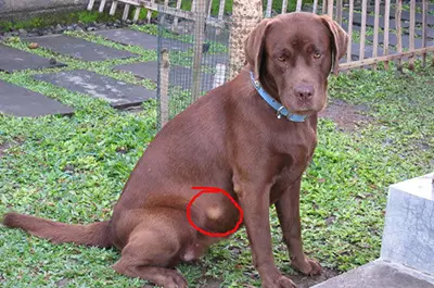
Source: https://s3.amazonaws.com/amcny/wp-content/uploads/2018/03/lipoma.jpg
Wart
Warts are caused by infection with a virus (called papilloma virus) that is transmitted by direct contact with an infected animal or contaminated objects like bedding or toys. They are often found around the mouths of young dogs. They tend to go away by themselves, but older dogs might need surgery to remove them.
Abscess
An abscess is a local accumulation or pocket of pus that can occur in any part of the body. Usually it is a sign of infection and the area surrounding the abscess is swollen and inflamed. A skin abscess is called a boil. It can be caused by an infection or a bite from an insect or other creature.
Mass
A mass is simply a lump in the body. Typically, any growth bigger than 3 centimetres in diameter is called a mass. It may be caused by the abnormal growth of cells, hormonal changes, or an immune reaction.
Tumour
A tumour is a mass lesion that is characterised by an abnormal mass of cells, lump of tissue or abnormal growth that may be benign or malignant. Tumours differ from other mass lesions in that the cells in tumours are abnormal in some way, for example, the appearance or size of the cells, the number of cells, or the regression of the cells’ physical characteristics toward a more primitive or undifferentiated type.
Neoplasm
A neoplasm is similar to a tumour and refers to a tumour that originates from a single cell and undergoes multiple duplications. These terms are often used synonymously in the literature. Sometimes “tumour” and “mass” are used to describe the actual swelling or other physical appearance of a neoplasm.
Mass lesions can occur in any part of the animal’s body, including:
- The skin
- The breasts (mammary glands)
- The head and neck
- The testicles
- The abdomen
- The bones
- The lungs
- The urinary tract
- The immune system
Cost of mass lesion treatment for dogs and cats
Mass lesion was the fifth most common health issue experienced by dogs and the tenth most common health issue experienced by cats in 2024, according to PetSure claims data.
Claims data for mass lesion |
Average cost of treatmentØ |
Highest cost of treatmentØ |
| Dogs | $961 | $31,195 |
| Cats | $1,486 | $34,688 |
ØBased on PetSure claims data, 2024 calendar year. Reimbursement for these claims under a pet insurance policy would be subject to limits, such as annual benefit limits or sub-limits, benefit percentage, applicable waiting periods and any applicable excess. Cover is subject to the policy terms and conditions. You should consider the relevant Product Disclosure Statement or policy wording available from the relevant provider. Please note that values calculated are based on all claims for that condition and medically related conditions in each calendar year.
Because it is difficult to predict the costs of veterinary care, it can help to have measures in place to help prepare for the unexpected. Pet insurance can help by covering a portion of the eligible vet bill if the unexpected does happen.
Get a quote for 2 months free pet insurance for your puppy or kitten in their first year.
Benign mass lesions
In many cases, the cells of the mass lesion are normal in appearance; such masses are usually benign. Benign growths tend to stay encapsulated in the place of origin and do not metastasise (spread). However, they can be aggressive and grow to huge proportions, pushing aside the surrounding tissues without invading them.
Benign mass lesions commonly found on dogs and cats include lipomas, sebaceous (skin) cysts, warts, infected hair follicles, and hematomas (blood blisters). While generally less cause of concern to owners, benign mass lesions can still create pain and discomfort for the animal. The veterinarian will advise which should simply be monitored, and which should be treated.
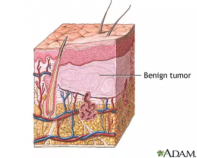
Source: https://medlineplus.gov/ency/images/ency/fullsize/9102.jpg
Malignant mass lesions
Other mass lesions are composed of cells that appear different from normal cells in size, shape, and structure; they usually belong to growths that are malignant. Their cells may be bizarre in form or may be arranged in a distorted manner. They are not encapsulated and therefore can invade the surrounding tissues. Typically, the less differentiated the cells are, the more quickly the tumour can be expected to grow.
Malignant mass lesions on dogs and cats tend to spread rapidly and can metastasise to other areas of the body. Examples of these include:
Soft tissue sarcomas
These typically occur as slowly growing masses anywhere in the body. When it occurs outside of a body cavity, a non-painful mass may be the only abnormality noted. When arising within the chest or abdominal cavity, it may be difficult to identify the tumour until it is very large.
Mammary tumours
Mammary tumours can be small, simple nodules or large, aggressive, metastatic growths. The tumours can occur in one or more glands. Mammary tumours are among the most common type of mass lesions found in female dogs, occurring most frequently in intact (entire) females.
Malignant melanoma
Most malignant melanomas occur in the oral cavity and the mucous membranes of the skin. The tumours can be highly pigmented or lack pigment and may invade deeply into the sub-cutaneous tissue; they grow rapidly and can be fatal. They usually metastasise via lymphatics to regional lymph nodes and lungs. They can also spread to other unusual parts of the body like the brain, heart and spleen.
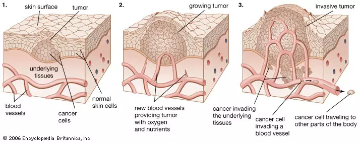
Source: https://cdn.britannica.com/64/91764-004-0294A28A.jpg
Symptoms of mass lesion in dogs and cats
The specific symptoms of a mass lesion vary depending on which part of the body is involved. Those near the skin can often be seen and/or felt. However, deeply placed tumours will not be visible and may not be palpable. The visual appearance of a mass lesion is highly variable, and may include:
- Any lump or bump or abnormal patch of skin
- An abnormal swelling that persists or continues to grow
- A hardened crack or fissure
- A wart-like projection
- Redness, bruising and fluid build-up (oedema)
- Sores that do not heal
- Ulcerations
- Fluctuations in size
Mass lesions that are most concerning tend to have the following characteristics:
- Grow fast
- Feel firm
- Change size or shape over a few weeks or months
- Ooze or break open
- Are firm and tightly fixed in place
- Are abnormally coloured
Generalised symptoms associated with a mass lesion, depending on its type, size and location, may include:
- Weight loss
- Loss of appetite
- Bleeding or discharge from any bodily opening
- Offensive odour
- Difficulty eating or swallowing
- Hesitation to exercise or loss of stamina
- Persistent lameness or stiffness
- Difficulty breathing
- Difficulty urinating or defecating
- Listlessness, lethargy
- Swollen abdomen
- Pale gums
- Vomiting
- Diarrhoea
- Seizures
Pain is a variable symptom with mass lesions. It is most often caused by pressure from the growing mass on adjacent nerves. In their early stages, masses tend to be painless, and those that grow to a large size without interfering with local functions may remain painless. Most malignant masses will eventually cause pain by the direct invasion of nerves or the destruction of bone.
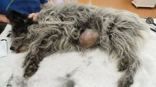
Source: https://i0.wp.com/farm9.staticflickr.com/8213/8418442575_589610490d.jpg
Causes of mass lesion in dogs and cats
A mass lesion is typically a mass of abnormal tissue that arises without obvious cause from pre-existing body cells. The underlying factors instigating the development of growths such as fluid-filled cysts, fatty tumours and many skin tumours tend to be unclear, although some are caused by a wound, previous trauma, hormonal changes, an immune reaction or the aging process.
The cause of a cancerous mass lesion in an individual animal is often unknown and may include genetic predispositions or exposure to several factors that disrupt normal cellular behaviour and induce changes in cells. Many different mechanisms give rise to malignancy, which makes diagnosis and treatment challenging.
There are a number of contributing factors that can increase the likelihood that cells will multiply, thereby raising the chances that cells will mutate during the division process and become cancerous, such as:
- Sun damage to the skin
- Areas of the body with little or no hair
- Light-coloured or thin coats
- Trauma, inflammation, irritants on the skin or compulsive licking of a spot on the skin
- Genetic factors in some breeds; for e.g. a mast cell tumour is the most common skin cancer in dogs and is most often found in Boxers, Boston Terriers, Labradors, Beagles, and Schnauzers
- The papilloma virus may be connected to the development of squamous cell tumours in certain dogs
- Age; older dogs and cats are more at risk for neoplasia than young animals
Unsure how serious it is?
Bow Wow Meow policyholders can get access to trusted vet care anytime, anywhere, at no additional cost. Connect to an experienced Australian registered vet via video call, 24/7. Whether it’s providing vet advice, setting up at-home treatment plans, or confirming if you need to visit a vet in person, you can get help when you need it.
Find out more about our pet insurance cover options.
How is mass lesion in dogs and cats diagnosed?
If the owner becomes aware of a mass lesion, he or she will need to provide the following history to the veterinarian:
- The mass or lump appeared suddenly, or it has been there a while
- The mass has retained the same consistency, or its appearance or has recently changed
- It seems to separate from the underlying tissue, or it seems fixed in place
- There appears to be only one lump, or there are multiple bumps
- The animal has had any changes in behaviour such as loss of appetite, weight loss, vomiting, diarrhoea, lethargy, or a dramatic change in overall attitude
The veterinarian will perform a physical examination, which includes a thorough head-to-toe observation and palpitation of the affected area. The look and feel of a mass can provide important information about it.
In addition, the veterinarian will usually recommend blood and urine tests as well as imaging studies such as radiographs, ultrasound, CT or MRI (magnetic resonance imaging) studies to define the extent of the mass. If there is a possibility of the mass being cancerous, the veterinarian will recommend a biopsy to confirm the diagnosis and the source of the malignancy.
Tissue biopsy
A tissue biopsy is performed by removing a small portion of the mass and sending it to a pathologist for microscopic examination. Biopsies can be obtained with a large needle, or with a small surgical procedure, usually under sedation or general anaesthesia. The mass may be removed in its entirety or a small piece may be excised.
Fine needle aspiration
Many lumps can be analysed via a needle biopsy rather than by tissue biopsy. Fine needle aspiration with cytology may be performed to differentiate between benign and malignant tumours. A sterile needle is inserted into the mass and the syringe plunger is used to “vacuum” in cells from the lump. The collected cells are smeared onto a glass slide for examination under a microscope.
Impression Smears
A glass microscope slide is pressed against the raw surface of the mass lesion. The collected cells are dried and sent to a pathologist for staining and diagnosis.
CT Scans or MRIs
These procedures are usually reserved for internal organ analysis. If a superficial malignant tumour is diagnosed, however, a CT scan or MRI can be helpful in determining if metastasis to deeper areas of the body has occurred.
Radiography and Ultrasonography
X-ray and ultrasound evaluations are generally reserved for collecting evidence of internal masses or metastases. Plain x-rays may reveal a large internal mass, but generally masses can be difficult to identify on plain x-rays. Contrast radiographs may be useful at times when ultrasonography fails to take clear images. X-rays of the lungs are also used to evaluate for metastatic disease, prior to treatment.
Ultrasounds are generally much more effective than radiographs in identifying an internal mass. Ultrasound is important for identifying the location of the mass, evaluating other metastatic sites, improving staging, guiding needle aspiration, biopsy and assisting in charting the proper course of treatment.
Prognosis
Whether the mass lesion is an inflamed cyst that needs draining or a tumour that requires surgical removal, discovering it early in its development and having it promptly checked by the veterinarian can make treatment easier and increase the odds of a favourable outcome. Any mass lesion, even if benign, may produce death by local effects by virtue of its size and/or location.
Malignancy refers to the ability of a tumour ultimately to cause death and is therefore a key factor in determining the prognosis. The chances of survival in cases of malignancy depends on many factors, including the location and size of the mass lesion and whether the cancer has metastasised (spread). For example, in the case of mast cell tumours, surgical removal those that have not metastasised may result in a cure. In the case of mammary tumours, if they are less than 3 cm in size the prognosis is quite positive; however, for tumours more than 1.5 inches in diameter the chances of survival are poor. In cases where it has metastasised to the lymph nodes, prognosis is very poor.
Treatment for mass lesion in dogs and cats
Treatment for mass lesion depends on the underlying cause and may range from no intervention to aggressive management. In cases of malignant tumours, treatment depends upon the age and overall health of the animal as well as the type and stage of cancer. Each case needs to be evaluated based on its own circumstances, but treatment for mass lesions typically include one or more of the following:
Conservative management
In many cases, a mass lesion will not affect the dog or cat and there will be no discomfort or interference with its daily life. In these cases, where the mass has remained the same size, the veterinarian may advise to leave it in place if it is not bothering the animal. This is especially true for animals that are unfriendly or difficult to manage in the surgery or that have complicating health conditions that make them poor candidates for surgical treatments.
If the veterinarian recommends leaving the mass lesion alone, it will be important to monitor it for any changes. In some cases, masses such as lipomas can grow extremely large, restrict the animal’s movement and become uncomfortable and bothersome. In this likelihood, surgical removal will be necessary.
Non-steroidal anti-inflammatory drugs may be prescribed if the animal is in pain. Stronger analgesic drugs or drug combinations may be required over time.
Minor procedures may be useful in the treatment of some mass lesions, for example:
Draining a cyst – where a biopsy of a cyst has come back with no indication of cancerous cells, the vet may choose to drain the cyst. This is typically done by placing a needle in the cyst and withdrawing the fluid. This is not painful and does not require sedation.
Lancing a cyst – where the fluid of a cyst is too dense, the vet may need to lance the cyst. This will require an anaesthetics that affects the localised area of the cyst only. The vet will then use a sharp blade to cut a small incision into the skin, allowing the contents of the cyst to drain. Stitches are not used to give the cyst the best opportunity to continue to express the fluid.
Surgical treatment
- Benign mass lesions
Surgical excision can be performed to remove a nuisance or dangerous lump. Even if a mass is benign, if it is causing a blockage or impeding mobility, it should be removed. Lipomas, for example, do not usually pose any serious health threat, but removal is recommended if they grow too large and limit the animal’s mobility, or the animal scratches or bites at them.
When a mass is small, it may not be much more invasive to remove it completely than to take a biopsy first. The smaller the mass, the easier it is to remove, meaning a less invasive surgery is required. If a biopsy reveals that a mass is benign, then less of the healthy tissue surrounding the mass will need to be removed.
Sometimes small skin growths or warts are removed as an “outpatient” procedure.
Other benign growths are removed under general anaesthesia and require more intensive post-op care. In some cases, cysts continue to recur and grow. These cysts will require more complicated surgery under anaesthesia in order to completely excise the follicle and the cyst wall.
If it becomes too big, even a benign mass can be very challenging to remove. It will cause more discomfort and will cost more in vet care. In some cases, even a benign mass may require an amputation because it has become too large.
- Malignant mass lesions
Surgery is the most commonly used treatment for cancerous mass lesions. There are various surgical options depending upon the size of the tumours, fixation to the surrounding tissue and the number of lesions. If a biopsy reveals that the mass is cancerous, the veterinarian will want to remove as much of it as possible, including adequate surgical margins (removal of healthy tissue adjoining the mass in every direction). Depending on the location of the mass lesion, this may be difficult, if not impossible, in some areas where there is little extra tissue, such as the foot or the leg, and amputation may be required.
Mast cell tumours: Surgery is the ideal treatment for mast cell tumours, provided the mass can be completely removed with adequate margins and it has not already spread. Depending on the size and location of the tumour, this may be performed with or without radiation therapy. Based on the grade of the cancer and the degree to which it has spread, chemotherapy and/or steroids may be used, as well.
Malignant melanomas: Surgery is the most common treatment for malignant melanomas. There are various surgical options depending upon the size of the tumour, fixation to the surrounding tissue and the number of lesions. These include lumpectomy, mammectomy, radical mastectomy, unilateral or bilateral mastectomy.
Squamous cell carcinomas: These can often be removed surgically, with no need for radiation or chemotherapy. If the tumours occur in inoperable locations, photodynamic therapy and the use of a non-steroidal anti-inflammatory drug (NSAID) may be beneficial.

Chemotherapy
The aim of chemotherapy is to eradicate a malignant tumour while maintaining quality of life. Chemotherapy medications are highly toxic to rapidly dividing cells and are an important mode of treatment for cancers that have spread to multiple locations within the body. It is often utilised after a cancerous mass has been removed via surgery but has a high likelihood of metastasising or has already done so.
Radiation
For inoperable and invasive tumours that do not have well-defined borders or are in a location that makes surgery difficult, radiation therapy may be useful. Some animals achieve a complete regression of the tumour. Radiation therapy is also beneficial in treatment of incompletely removed tumours. If a malignant melanoma cannot be removed in its entirety or if it has spread to nearby lymph nodes, radiation is commonly used. It may be used alone or in combination with other treatments, typically surgery and/or chemotherapy. Sometimes radiation therapy is used intra-operatively as well as post operatively to reduce the chances of recurrence.
Experimental
Some cancers, such as canine melanoma, have been found to be highly resistant to chemotherapy, while others may occur in inoperable locations. In these cases, alternative treatments may be sought. Emerging techniques such as gene therapy, photodynamic therapy and immunotherapy are showing promise to combat some types of tumours in dogs and cats.
Immunotherapy is slowly emerging as a potential alternative for controlling the growth of tumour cells. A therapeutic vaccine has been launched, but its availability is restricted. It is mainly indicated for the treatment of dogs with oral melanomas.
In summary
 A mass lesion is an abnormal lump in the body, such as a cyst, wart, abscess, lipoma or tumour. It may be benign (not cancerous) or malignant (cancerous). Mass lesions can occur on any part of the body. Many occur on or under the skin, but they can also develop internally, for example on the organs and bones. They may be caused by the abnormal growth of cells, an infection, hormonal changes, an immune reaction or it may have no definable cause.
A common technique when diagnosing a mass lesion is to take a tissue biopsy or fine needle aspirate so that the cells can be microscopically examined. Surgical removal is often the treatment of choice, even where the mass lesion is benign. Some mass lesions are very aggressive and can rapidly increase in size. If it becomes too big, even a benign mass can be very challenging to remove. Other treatment options include no intervention, conservative management, chemotherapy and radiation.
More information
- http://www.pethealthnetwork.com/dog-health/dog-diseases-conditions-a-z/neoplasia-dogs
- http://www.pethealthnetwork.com/dog-health/dog-surgery-a-z/what-a-biopsy-and-when-might-your-dog-need-it
- http://www.pethealthnetwork.com/dog-health/dog-diseases-conditions-a-z/lipomas-also-known-lumps-and-bumps
- https://www.petmd.com/dog/general-health/evr_dg_lumps_and_bumps
- https://www.petmd.com/dog/conditions/8-types-dog-tumors-and-how-treat-them
Bow Wow Meow Pet Insurance can help protect you and your pet should an unexpected trip to your vet occur.
- Find out more about our dog insurance options
- Find out more about our cat insurance options
- Get an instant online pet insurance quote
Bow Wow Meow is proud to have been awarded winner of Canstar’s ‘Most Satisfied Customers’ Award in the Pet Insurance category for both 2024 and 2025!
Bow Wow Meow is proud to be the only Pet Insurer that has been awarded Product Review’s Top Rated Pet Insurance every year from 2018 to 2026! This is based on 3,193 independent customer reviews (as at 09/04/2026), with an overall rating of 4.5 stars.

We also have the following ratings on other review platforms:
Google Review rating of 4.5 stars (based on 1,358 reviews)
Trust Pilot rating of 4.6 stars (based on 555 reviews)
Bow Wow Meow has been chosen as Winner of 4 categories in the 2026 Finder Pet Insurance Customer Satisfaction Awards!


– Most Trusted Pet Insurance
– Easiest to Claim Pet Insurance
– Most Recommended Pet Insurance
– Legendary Service Pet Insurance
We also received Highly Commended Awards for Loved Brand Pet Insurance and Best Value Pet Insurance.
Bow Wow Meow is an Australian-owned and operated pet insurance provider. Our focus is purely on pets, and we are proud to have helped provide peace of mind to over 200,000 Aussie pet owners since 2008.