
Skeletal abnormalities in dogs and cats: signs, causes and treatment


Summary
Skeletal abnormalities are conditions that affect the normal growth, shape, strength or function of a dog’s or cat’s bones and joints. They may be inherited, develop while a young animal is growing, or occur later because of injury, infection, nutritional or metabolic disease, degeneration or cancer. Common signs include limping, pain, stiffness, swelling, reduced mobility and visible limb or spinal deformity. Any persistent lameness or sudden inability to bear weight should be assessed by a vet. Treatment may involve surgery, physiotherapy, weight management and pain control. Prognosis varies depending on underlying disease.
What are skeletal abnormalities in dogs and cats?
Skeletal abnormalities are conditions that affect the normal development, shape, strength or function of the bones and joints. Some are congenital, meaning they are present at birth, while others develop as a puppy or kitten grows. Skeletal problems can also be acquired later in life because of injury, infection, nutritional or metabolic disease, age-related degeneration or cancer.
These disorders may affect a single bone, several bones, a growth plate, the cartilage within a joint or the spine. Depending on the cause and severity, they may result in pain, lameness, swelling, reduced movement or visible deformity.
Expert advice from Dr Felicia:
Not every limp is caused by a skeletal disorder. Muscles, tendons, ligaments, joints and nerves can also cause pain or abnormal movement, so a veterinary examination is needed to locate the source of the problem.
Common skeletal and bone disorders
Skeletal conditions range from traumatic injuries to inherited and developmental diseases. Common or clinically important examples include:
Bone fractures and traumatic injuries
Trauma to bones is probably the most common skeletal problem seen in dogs and cats. Broken bones (“fractures”) are often caused by car accidents or falls from a height. There are several types of fractures that can occur, including single or multiple and open (also called compound) or closed. Fractures most commonly sustained by dogs and cats are to the jaw, pelvis, and the fore- or hindlimbs.
Learn more about fracture of the skull, fracture of the forelimbs and fracture of the hindlimbs.
Hip dysplasia
Abnormal development and looseness of the hip joint, which can lead to pain, reduced mobility and osteoarthritis. Hip dysplasia occurs primarily in large breed dogs and is relatively rare in cats, although approximately 18% of Maine Coon cats are reported to suffer from it. Learn more about hip dysplasia in dogs and cats.
Elbow dysplasia
Several inherited conditions cause degenerative joint disease in the elbow, affecting elbow joint development. It is mostly seen in large and giant breed dogs and can result in lameness in one forelimb or a stiff gait when both elbows are affected. Dogs are usually affected around 6-12 months of age.
Osteochondrosis and osteochondrosis dissecans (OCD)
Developmental disorders in which cartilage within a growing joint does not mature normally. A weakened piece of cartilage may crack or form a flap, causing pain, inflammation and lameness. Although OCD has been seen in cats and small dogs, it occurs primarily in young, growing, dogs of large and giant breed and less than one year of age.
Hypertrophic osteodystrophy (HOD) and panosteitis:
Painful developmental bone conditions seen primarily in young, rapidly growing dogs (i.e. large and giant breeds). HOD affects the areas of bone close to the growth plates, while panosteitis, also known as “growing pains”, affects the long bones and may cause lameness that shifts between legs.

Angular limb deformities
Abnormal growth or alignment of one or more limb bones. These may be inherited or result from damage to a growth plate while an animal is young.
Osteochondrodysplasia
A group of inherited disorders affecting cartilage and bone development. Scottish Fold osteochondrodysplasia is one well-known example in cats. Due to a genetic mutation, bones do not grow to the normal size, based on what is expected of the breed. The result is abnormally short limbs, a condition called dwarfism.

Osteomyelitis
Inflammation and infection of bone or bone marrow, usually caused by a bacterial infection. It can occur following a traumatic injury (where there is bone damage and/or an open wound), or post-operatively. Infection can spread from the bone or bone marrow to the bloodstream.
Bone tumours
Osteosarcoma is the most common primary bone tumour in dogs and cats. It is typically aggressive in dogs and frequently spreads to other parts of the body, particularly the lungs. Feline osteosarcoma generally behaves less aggressively than the disease in dogs.
Signs and symptoms of skeletal abnormalies
While lameness is often the primary sign that may indicate a skeletal abnormality, symptoms can vary depending on the condition, for example:
Hip dysplasia – lameness, limping, pain, difficulty moving from lying to standing, a bunny-hopping gait in the rear legs, a reluctance to walk, and stiffness in the rear legs; in mild cases there may be no visible symptoms
Elbow dysplasia – lameness in forelimbs, reluctance to jump down, stiff gait when both forelimbs are affected.
Osteochondrosis dissecans (OCD) – pain (varying from mild and intermittent to severe and constant), limping in one or both hind limbs, trouble upon rising or appearing stiff when walking; a characteristic “slinky” gait has been described for some hind limb OCD animal
Hypertrophic osteodystrophy (HOD) – swelling and pain of the bone just above the joints, pain in the joints, fever and loss of appetite
Multiple cartilaginous exostoses (MCE) – lameness and discomfort
Osteogenesis Imperfecta – pain, loose joints, difficulty walking, spontaneous fractures, reduced bone mass, stunted growth, muscle atrophy
Osteochondrodysplasia and Achondroplasia – poor growth, abnormal bone shape, larger head than normal, shorter jaw and nose, crooked teeth, bowed forelimbs, skew spine, enlarged joints
Osteomyelitis (inflammation of bone) – lameness and pain, red, swollen and/or pus-filled sores (ulcers) at the wound site, fever, lethargy, persistent decreased appetite and depression
Osteochondromatosis (Multiple Exostoses) – there may be no signs, or lameness or pain may develop
Panosteitis (inflammation in the marrow cavity of the long bones) – difficulty in moving, pain in the bone or limb, lameness, fever, loss of appetite and lethargy
Bone tumour / cancer – symptoms depend on the site of the tumour, including lameness where the tumour is in a limb, bone swelling, and fractures of the bone that are not caused by injury
When should you see a vet?
Arrange a veterinary appointment if your dog or cat has persistent or recurring limping, stiffness, difficulty getting up, reduced activity, reluctance to jump or climb stairs, pain when touched, swelling around a bone or joint, or a limb that appears bowed or unusually shaped.
Seek urgent veterinary care if your pet:
- suddenly cannot place weight on a limb
- has been hit by a vehicle or experienced a serious fall
- has severe pain, marked swelling or an obviously deformed limb
- has an open wound near a suspected fracture or visible bone
- is dragging a limb or has suddenly lost coordination
- develops fever, lethargy or loss of appetite with bone or joint pain
- develops a fracture without an obvious injury
Avoid repeatedly manipulating a painful limb. Keep your pet as still as possible and contact your vet for advice about safe transport.
Expert advice from Dr Felicia:
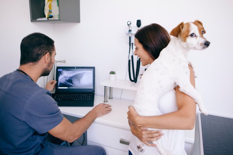
While a physical examination and history are likely to give helpful clues to the area affected, a radiograph is usually required to visualise abnormalities or injury to the bone and confirm the diagnosis.
What causes skeletal abnormalities?
The causes of skeletal abnormalities can be hereditary (or breed-related / genetic). developmental, nutritional, infectious, traumatic, neoplastic (cancer) secondary to other diseases or of unknown origin. Some skeletal abnormalities in dogs and cats are thought to be caused by a combination of these factors. Osteochondrosis dissecans (OCD), for example, has a causal pathway that includes genetics, rapid growth, excessive dietary calcium, and trauma.
Inherited and congenital conditions
A hereditary or genetic condition is one that is passed down from one or both parents and/or is more prevalent in a particular breed. Osteogenesis imperfecta (brittle bones) and polydactyly (having extra toes) are examples of hereditary disorders. Some hereditary diseases are present at birth while others become manifest during growth or later in life (i.e., acquired hereditary diseases).
Often more than one gene is involved in hereditary skeletal abnormalities, and in many of these conditions environmental factors also play a role. For instance, hip dysplasia is a common hereditary disorder with multiple genes involved, occurring in many large breeds of dogs such as Golden Retrievers, Labradors, Mastiffs and German Shepherds; however, the development and progression of hip dysplasia is somewhat influenced by environmental conditions such as obesity, rapid weight gain and poor nutrition.
The risk of some inherited skeletal disorders can be reduced through responsible breeding practices. These may include breed-specific screening, genetic testing where a validated test is available, assessment of relatives and avoiding breeding animals affected by serious inherited disease. Breeders should seek advice from their veterinarian and relevant breed health programs.
Developmental disorders
A developmental condition is one that occurs during the animal’s development, either in utero or later in life. Developmental bone disorders appear in young animals when the bones do not grow correctly. They may be congenital (present at birth) or occur as the animal grows.
Nutritional deficiencies in the mother’s diet during pregnancy can cause malformations in the skeletal system of the growing foetus. Hereditary breed characteristics can also cause developmental abnormalities, for example, Bulldogs, Pugs, Boston Terriers, Basset Hounds and Dachshunds are susceptible to abnormal development of the bones of the foreleg (radius and ulna), resulting in lameness.
Trauma
Trauma to bones is regarded as the most common cause of skeletal disorders in dogs and cats, especially in animals allowed to roam free. Trauma includes falls from a height, automobile accidents, fights and even gunshot wounds.
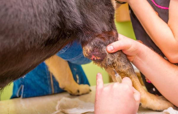
Infection
The most common cause of inflammation of the bone or bone marrow is bacterial infection, as in the case of osteomyelitis. Bacterial contamination of a wound such as a torn nail, fracture, bite wound or laceration near a bone can spread to the bone. Infections that cause bone tissue to break down and die can lead to bone disorders.
Systemic infections (infections occurring in other areas of the body) can also reach the bone or marrow through the bloodstream; for example, when osteochondromatosis occurs in older cats, it is believed to be caused by infection with the feline leukaemia virus. Less common causes of bone infections are fungal infection or the presence of surgical implants, such as a prosthetic joint or bone plates and pins.
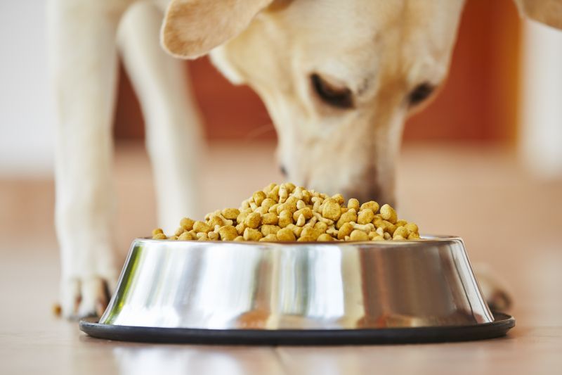
Nutritional factors
When animals are not fed the right diet for their breed and/ or weight and/or age, nutrition-related bone conditions can occur. For example, nutrition is suspected to play a role in osteochondrosis and hypertrophic osteodystrophy. Reduced bone mass, bone deformities, bony growths, fractures, lameness and loose teeth are all conditions that can result from nutritional deficiencies.
Mineral imbalances in the diet, particularly of trace minerals such as copper, zinc, and magnesium, are a common dietary cause of bone defects. An excess or deficiency of vitamins, particularly vitamins A and D, can also influence bone growth and development. A deficiency in vitamin D in the diet, or low conversion of vitamin D in the body from a lack of exposure to sunlight, can affect the development of bone.
An excess of protein in the diet can cause an imbalance of calcium and phosphorus in the body, which can develop into nutritional disorders affecting the bones. A calcium deficiency or imbalance in the diet can cause rickets, a severe weakening of the bone that arises most often in young dogs fed an all-meat diet. In puppies it causes lameness, deformities and fractures of the bone.

Metabolic disease
Some disorders that cause abnormalities in the circulating levels of calcium, phosphorous and certain vitamins can adversely affect bones. Chronic kidney failure affects bone by altering the amount of phosphorus and vitamin D in the body. The bones become soft, thin, and weak.
Metabolic diseases result from too much or too little of a particular hormone or other substance in the body. Panosteitis, osteochondrosis, and hypertrophic osteodystrophy are the three most common metabolic bone disorders seen in dogs.
Bone cancer
Skeletal tumours can arise within the tissues of the bone (primary cancer), or can invade bones from the surrounding soft tissues, or spread to the bone from a primary location in another part of the body (metastasis). The most common primary bone tumour is osteosarcoma of the radius, humerus, femur, or tibia.
Rarely, tumours can occur at sites of previous bone damage, including fractures, orthopaedic implants (used for fracture repair or joint replacement), radiation therapy, and bone diseases. Most primary bone tumours, particularly osteosarcoma, develop spontaneously with no apparent predisposing cause. There appears to be a genetic predisposition to developing osteosarcoma in Scottish Deerhounds and this tumour also occurs frequently in other large breed dogs, particularly the Rottweiler. Large or giant, and particularly tall, dogs are at a greater risk for the development of osteosarcoma compared to the general dog population. Learn more about cancer in dogs.
Unknown origin
The exact causes of some skeletal abnormalities are unknown, although a combination of the above factors may play a role. For example, some factors thought to play a role in the origin of panosteitis include genetics (in German Shepherds), stress, infection, or the body’s own metabolic and immune responses.
Expert advice from Dr Felicia:
As pet owners most of these causes are far out of our control. What we can do is aim to provide good nutrition, appropriate exercise and particularly for large breed dogs, consider what genetic testing has been performed on the parents of a puppy prior to purchase.
How are skeletal abnormalies diagnosed?
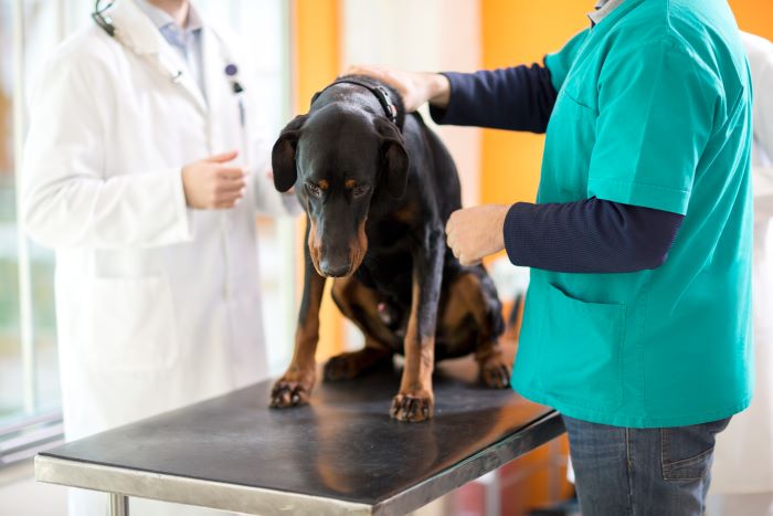
Diagnosis begins with a detailed history and physical examination. Your vet may ask when the signs began, whether there has been an accident or injury, whether the problem is getting worse and whether similar conditions have occurred in related animals.
During an orthopaedic examination, the vet may observe your pet standing, walking or trotting and then feel the limbs, bones, joints and spine for pain, swelling, instability, reduced range of motion or deformity.
Expert advice from Dr Felicia:
Lameness and pain doesn’t immediately localise itself to the bone. Soft tissue injury, joint problems and diseases affecting the nervous system can present similarly. Your vet’s examination will help to figure out whether your dog or cat’s issue has a skeletal cause.
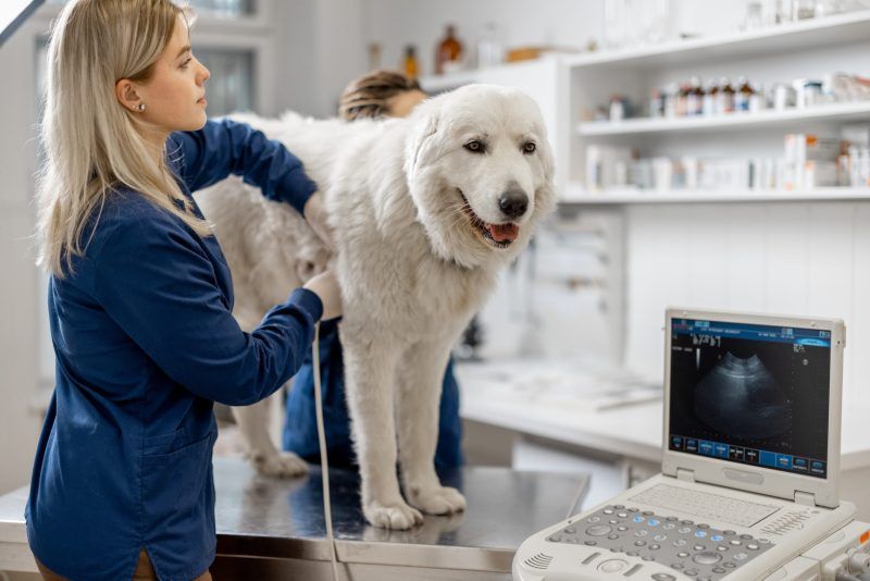
Radiography
X-rays are a crucial tool in evaluating the skeleton. Bone and some forms of cartilage show up very well on x-rays. Sedation or anaesthesia may be needed to position the animal safely and obtain clear images.
Expert advice from Dr Felicia:
Radiograph can demonstrate changes to the bone. These may be very distinctive in their characteristics or location depending on the disease. However for abnormal/rare diseases, to achieve a definitive diagnosis a biopsy may be required to evaluate the cells present in the abnormal/diseased bone.
Routine laboratory tests
A complete blood count, biochemistry profile and urinalysis are performed to assess general health and look for signs of underlying infection and/or abnormalities in circulating levels of calcium, phosphorus and vitamin D. These tests are also helpful to detect other underlying disorders that may affect the bones, such as kidney disease, anaemia and leukaemia.
Special laboratory tests
If an infection of the bone is suspected, samples of fluid or pus may be collected for analysis and bacterial or fungal cultures. Occasionally the levels of vitamin D and parathyroid hormones are measured in the blood.
Genetic tests
If the condition is identified as genetic (inherited), a buccal (cheek) swab will be taken for laboratory analysis to determine if the animal is a carrier of the gene.
Bone biopsy
Identification of the type of bone disease present may require a bone biopsy, where a core sample of the suspected tumour is obtained for analysis by a pathologist. This is especially recommended where the presentation is atypical, in cases of congenital and developmental bone diseases, osteomyelitis and bone tumours. In cases of tumours, a biopsy of the suspected tumour itself is taken; because different types of tumours respond differently to various treatments, a biopsy can help to determine the best treatment plan.
Advanced imaging techniques
CT (computed tomography) scans and MRI (magnetic resonance imaging) are very useful in examining bones and their adjacent soft tissues, such as cartilage, ligaments, and tendons. CT scans are recommended for many tumours of the axial skeleton as the cross-sectional and three-dimensional images provide better information than traditional radiography.
Chest x-rays
Chest x-rays may be taken to look for evidence of infection or tumours that have spread to the lungs, and to identify abnormalities in the ribs or vertebrae of the chest.
Treatment and management
Treatment depends on the type of skeletal abnormality, the bones or joints affected, the severity of the condition and the pet’s age and general health. Some problems can be managed with rest and medication, while others require surgery or specialist treatment.
Expert advice from Dr Felicia:
Considerations for treatment likely need to take into account the dog’s breed and activity level, lifestyle, finances and ongoing requirements for monitoring or treatment. In some scenarios pursuing treatment may be too stressful or not appropriate for the patient depending on the wider clinical picture. You can discuss your concerns with your vet to decide what makes the most sense for your pet.
Surgery
Surgical procedures vary depending on the presenting skeletal abnormality:
Tumours – for limb osteosarcoma in dogs, amputation is commonly recommended to remove the painful tumour and reduce the risk of a pathological fracture.
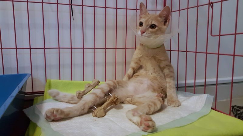
Fractures – surgery to stabilise a bone fracture includes the use of implants or other fixation material, such as plates, screws, wires and pins, depending on the location and severity of the fracture.
Osteochondrosis dissecans (OCD) – optimal treatment typically involves surgery to remove abnormal cartilage, performed by arthroscopy, where a small camera attached to a flexible tube, or endoscope, is inserted into the joint through a small incision, or arthrotomy, an open-joint procedure.
Hip dysplasia – there are four main surgeries that may be recommended, depending on the animal’s size and age: triple pelvic osteotomy (TPO), juvenile pubic symphysiodesis (JPS), and total hip replacement (THR). A salvage procedure that may be suitable for smaller patients is a femoral head ostectomy (FHO).
Amputation – if there is too much damaged bone or tissue damage, amputation of a digit, tail, or limb may be the recommended treatment option, or the only effective strategy for saving the animal’s life.
Because it is difficult to predict the costs of veterinary care, it can help to have measures in place to help prepare for the unexpected. Pet insurance can help by covering a portion of the eligible vet bill if the unexpected does happen.
Get a quick quote
Conservative management
Conservative treatment options include medications, exercise restriction (until the signs completely subside), and appropriate weight and nutritional management.
Medication
Relieving pain is an important component of treatment for lame animals and may allow faster recovery. Pain medication can also help animals with chronic conditions, such as osteoarthritis. For example, your vet may prescribe appropriate nonsteroidal anti-inflammatory drugs (NSAIDS) to reduce swelling and inflammation, and/or pain-relieving medications to reduce the severity of the pain caused by bone abnormalities.
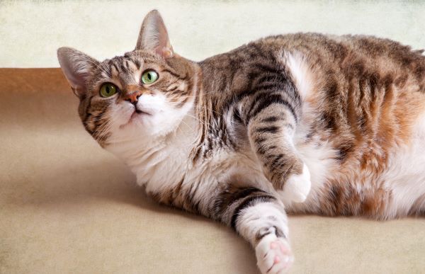
Weight control
Weight control is an important aspect of recovery and ongoing management to decrease the pressure applied to the affected limb or joint. The risk of weight gain needs to be kept in mind where physical mobility is reduced, whether there is a reduction of exercise during recovery or as a long-term consequence of the condition.
Nutrition
Ensuring that a dog or cat has the proper nutrition required for strong bones is critical for optimal health. Good nutrition is particularly important during the growing years, especially for animals of susceptible breeds, to decrease the likelihood of some skeletal abnormalities. Reduced bone mass, bone deformities, bony growths, fractures, and loose teeth are all conditions that can result from nutritional deficiencies.
Conditions requiring long-term supportive management
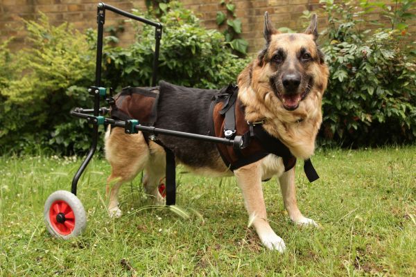
Some inherited or progressive skeletal disorders cannot be cured, but pain relief, environmental changes, rehabilitation and other supportive treatments may help maintain mobility and quality of life. Some animals learn to compensate for their disability and manage to live relatively normal lives. However, in severe cases, euthanasia may be recommended on welfare grounds.
Other treatment options may include, where appropriate:
- Physiotherapy, acupuncture or massage (passive joint motion) to decrease joint stiffness and help maintain muscle integrity
- Chemotherapy, usually following surgery, particularly in cases of osteosarcomas
- Hospitalisation with bed rest and frequent turning
- Intravenous fluid therapy, if the animal is dehydrated
- Irrigation, flushing, cleaning and draining of any open wound
- Removal of dead, damaged, or infected tissue
- Removal of loose implants
- Bone grafting
- Stem cell therapy
- Palliative treatment, in cases of malignant tumours, to provide pain control and improve comfort for the remaining lifespan
Can skeletal abnormalities be prevented?
Not every skeletal abnormality can be prevented, particularly when a condition is inherited or congenital. However, you can help support healthy bone and joint development by feeding a complete and balanced diet suitable for your pet’s life stage, avoiding unnecessary calcium or vitamin supplements and maintaining a healthy body weight.
Puppies, particularly large- and giant-breed puppies, should grow at a steady, controlled rate rather than being overfed. Appropriate exercise, safe surroundings and prompt treatment of injuries can also reduce the risk of trauma-related and developmental problems. Responsible breeding and breed-specific health screening can help reduce the incidence of some inherited skeletal disorders.
Prognosis
The outlook for a pet with a skeletal abnormality depends on the underlying condition, its severity, the pet’s age and general health, and how early treatment begins. Some fractures and developmental problems can be corrected successfully, while other conditions require lifelong management.
Panosteitis and many mild-to-moderate cases of hypertrophic osteodystrophy may improve as a young dog matures. In contrast, progressive inherited disorders and malignant bone tumours can have a more guarded prognosis. Your vet can provide a more individual outlook once the condition has been identified and staged
In summary
Dogs and cats can suffer from disorders that affect the skeletal system, resulting in lameness or bone deformities. Skeletal abnormalities can be developmental, genetic, infectious, nutritional, traumatic, or neoplastic in origin. In many cases there is an interplay of inherited breed characteristics with environmental and other factors. The degree of impairment depends on the specific problem and its severity but limping and pain are symptoms common to many of them.
X-rays are a crucial tool in diagnosing skeletal abnormalities. Treatment options range from medication to relieve pain and inflammation, to surgery, to no treatment. Supportive care and dietary changes may be recommended. Prognosis is varied, with some skeletal abnormalities having a much more optimistic outlook than others.
Bow Wow Meow Pet Insurance can help protect you and your pet should an unexpected trip to your vet occur.
- Find out more about our dog insurance options
- Find out more about our cat insurance options
- Get an instant online pet insurance quote
Bow Wow Meow is proud to have been awarded winner of Canstar’s ‘Most Satisfied Customers’ Award in the Pet Insurance category for both 2024 and 2025!
Bow Wow Meow is proud to be the only Pet Insurer that has been awarded Product Review’s Top Rated Pet Insurance every year from 2018 to 2026! This is based on 3,193 independent customer reviews (as at 09/04/2026), with an overall rating of 4.5 stars.

We also have the following ratings on other review platforms:
Google Review rating of 4.5 stars (based on 1,358 reviews)
Trust Pilot rating of 4.6 stars (based on 555 reviews)
Bow Wow Meow has been chosen as Winner of 4 categories in the 2026 Finder Pet Insurance Customer Satisfaction Awards!


– Most Trusted Pet Insurance
– Easiest to Claim Pet Insurance
– Most Recommended Pet Insurance
– Legendary Service Pet Insurance
We also received Highly Commended Awards for Loved Brand Pet Insurance and Best Value Pet Insurance.
Bow Wow Meow is an Australian-owned and operated pet insurance provider. Our focus is purely on pets, and we are proud to have helped provide peace of mind to over 200,000 Aussie pet owners since 2008.
Sources
“Developmental Osteopathies in Dogs and Cats”, MSD Veterinary Manual, https://www.msdvetmanual.com/musculoskeletal-system/osteopathies-in-small-animals/developmental-osteopathies-in-dogs-and-cats. Accessed 23 July 2026.
“Congenital and Inherited Anomalies of the Musculoskeletal System in Dogs and Cats”, MSD Veterinary Manual, https://www.msdvetmanual.com/musculoskeletal-system/congenital-and-inherited-anomalies-of-the-musculoskeletal-system/congenital-and-inherited-anomalies-of-the-musculoskeletal-system-in-dogs-and-cats. Accessed 23 July 2026.
“Osteochondrosis in Dogs”, MSD Veterinary Manual, https://www.msdvetmanual.com/musculoskeletal-system/arthropathies-and-related-disorders-in-small-animals/osteochondrosis-in-dogs. Accessed 23 July 2026.
“Osteomyelitis in Dogs and Cats”, MSD Veterinary Manual, https://www.msdvetmanual.com/musculoskeletal-system/osteopathies-in-small-animals/osteomyelitis-in-dogs-and-cats. Accessed 23 July 2026.
“Bone Tumors in Dogs and Cats”, MSD Veterinary Manual, https://www.msdvetmanual.com/musculoskeletal-system/osteopathies-in-small-animals/bone-tumors-in-dogs-and-cats. Accessed 23 July 2026.
“Bone Disorders in Dogs”, MSD Veterinary Manual, https://www.msdvetmanual.com/dog-owners/bone-joint-and-muscle-disorders-of-dogs/bone-disorders-in-dogs. Accessed 23 July 2026.
“Bone Disorders in Cats”, MSD Veterinary Manual, https://www.msdvetmanual.com/cat-owners/bone-joint-and-muscle-disorders-of-cats/bone-disorders-in-cats. Accessed 23 July 2026.
“Osteosarcoma in Dogs”, Cornell University College of Veterinary Medicine, https://www.vet.cornell.edu/departments-centers-and-institutes/riney-canine-health-center/canine-health-topics/osteosarcoma-dogs. Accessed 23 July 2026.