
Protein losing enteropathy (PLE) in dogs


Summary
Protein losing enteropathy occurs when inflammation, lymphoma or lymphaic disease in the gut causes protein loss into the intestines. Dogs may show diarrhoea, weight loss, swelling or fluid in the abdomen or even their chest. Diagnosis uses blood tests, ultrasound and biopsy where possible. Treatment targets the cause and includes low fat diets, immunosuppressants and B12 support. Close monitoring helps reduce relapse and improves long term outcomes, though prognosis is generally guarded for these patients.
What is protein losing enteropathy in dogs?
Protein-losing enteropathy (PLE) refers to an excessive loss of protein from the bloodstream in the intestinal tract. The word “enteropathy” means any disease of the intestinal system. Protein losing enteropathy is not a specific disease but a syndrome associated with several gastrointestinal (GI) diseases in dogs that result in a loss of protein from the intestines.
Normally, small amounts of protein leak out of the blood vessels as they pass through the intestines; these leaked plasma proteins are broken down and then reabsorbed by the body and used to make more proteins. With PLE, the mucosal layer that lines the inner surface of the intestine is compromised, allowing more protein-rich fluid to leak out than the body can replace. PLE can lead to a state of hypoproteinemia, a condition where there is an abnormally low level of protein in the blood.
There are several diseases that can damage the intestines sufficiently to cause this extra protein loss. Primary gastrointestinal disease, right-sided heart failure and diseases of the lymphatic system can all cause PLE.
PLE in dogs can affect any breed or age, but some breeds show a predisposition for certain disorders linked to PLE. These breeds include the Soft-coated Wheaten Terrier, Basenji, Yorkshire Terrier, Shar Pei and Norwegian Lundehund. However, no genetic predisposition has yet been proven.
Unsure how serious it is?
Bow Wow Meow policyholders can get access to trusted vet care anytime, anywhere, at no additional cost. Connect to an experienced Australian registered vet via video call, 24/7. Whether it’s providing vet advice, setting up at-home treatment plans, or confirming if you need to visit a vet in person, you can get help when you need it.
Find out more about our pet insurance cover options.
Symptoms of protein losing enteropathy in dogs
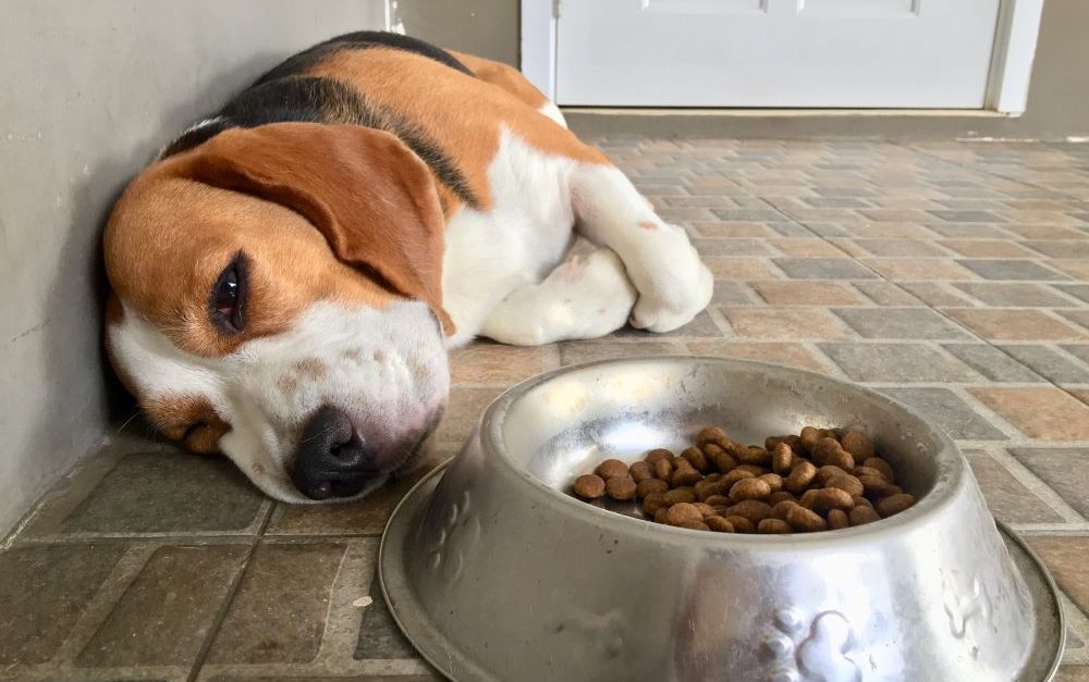
While symptoms of PLE in dogs may initially be subtle, if left untreated the disease can quickly progress to become severe and potentially life threatening. Weight loss, diarrhoea and vomiting are the most common clinical signs, but are not present in every case.
Expert advice from Dr Felicia:
Dogs with persistent diarrhoea or other gastrointestinal signs should have some blood tests and potentially imaging to ensure that there isn’t an underlying cause which needs to be addressed, and to identify if their disease is severe enough that their protein levels are falling below normal, as PLE is a serious disease that requires prompt identification and therapy to optimise outcomes.
Observable symptoms of PLE in dogs include:
- Bouts of diarrhoea
- Chronic diarrhoea
- Blood-tinged, mucoid, dark, sticky faeces (melaena)
- Weight loss
- Lack of energy (lethargy)
- Difficulty breathing (dyspnoea) (due to fluid accumulation in the chest cavity)
- Enlarged abdomen, pot-bellied appearance (due to fluid accumulation within the abdomen)
- Legs and feet may be puffy or swollen (oedema)
- Decreased appetite (anorexia)
- Being a “picky” eater
- Vomiting
Your veterinarian may detect additional symptoms on physical examination:
- Muscle wasting
- Malnutrition
- Thickened loops of intestine on palpation
- Fluid in the abdomen
- A heart murmur
- Dull lung sounds (from fluid on the lungs)
Causes of protein losing enteropathy in dogs
Excessive loss of protein can occur through the gastrointestinal (GI) tract as a result of many conditions but is most commonly caused in dogs by inflammatory bowel disease (inflammation of the intestines) and lymphoma (cancer of the lymphoid tissues in the GI tract).
Diseases and conditions of the GI tract that can lead to PLE include:
- Inflammatory bowel disease (IBD), including lymphocytic-plasmacytic enteritis, eosinophilic enteritis, granulomatous enteritis and histiocytic-ulcerative colitis
- Lymphatic diseases like intestinal lymphangiectasia (pathologic dilation of lymph vessels), lymphoma
- Parasitic enteritis from Giardia, hookworms and whipworms (more common in younger animals)
- Adverse food reactions
- Mechanical GI disease due to chronic foreign body irritation
- Intestinal cancer
- Gastrointestinal ulceration and haemorrhage (ulcers in the stomach or intestines)
- Chronic intussusception (a common cause in juvenile animals)
There are a number of cardiovascular diseases and conditions that can cause of PLE in dogs, including:
- Venous congestion and portal hypertension
- Posterior caval syndrome
- Right-sided congestive heart failure
How is protein losing enteropathy in dogs diagnosed?
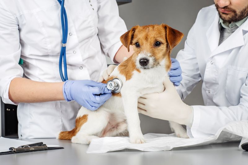
If you notice any of the symptoms listed above, consult your veterinarian immediately. Your vet will perform a physical examination and will ask you for a thorough history of your dog’s health and onset of symptoms you have observed.
In most cases, clinical signs, laboratory tests, imaging (usually ultrasound) with the endoscopic intestinal biopsies can lead to at least a provisional diagnosis of PLE and guide initial treatment. However, sometimes patients can be too unwell to have endoscopic biopsies performed and treatment is commenced with an incomplete picture, with the final diagnosis made after assessing the patient’s response to treatment.
As it is a syndrome rather than a disease, once PLE has been confirmed it is essential to find the underlying cause. Often this can only be accomplished by ruling out other possible causes. Although any intestinal disease that is severe enough to cause a net loss of protein can result in PLE, some diseases are commonly recognised to cause PLE, such as:
- Intestinal lymphangiectasia, characterised by dilation of the intestinal lymph vessels, believed to be the most common cause of PLE in dogs, can occur as a primary (congenital) or secondary (acquired) disease.
- Inflammatory bowel disease (IBD), defined as chronic inflammation of the bowel wall (e.g., lymphoplasmacytic enteritis), must be severe to result in intestinal protein loss.
- Lymphoma of the intestinal tract can cause hypoalbuminemia, abnormally low levels of albumin, a major blood protein, in 75% of dogs.
- Adenocarcinoma and intestinal tumours may cause PLE due to blood loss and mucosal ulceration.
- Gastroenteritis (from viral, fungal, or bacterial infections)
- Stomach ulcers
- Granulomatous infiltration of the intestines (secondary to fungal infections)
- Abnormalities of the intestines (from a chronic foreign body, intestinal parasites, an intussusception, haemorrhagic gastroenteritis, etc.)
Because it is difficult to predict the costs of veterinary care, it can help to have measures in place to help prepare for the unexpected. Pet insurance can help by covering a portion of the eligible vet bill if the unexpected does happen.
Get a quote for 2 months free pet insurance for your puppy or kitten in their first year.
Tests that are typically performed to diagnose PLE and/or rule out other diseases include:
Laboratory tests
Standard laboratory testing will be performed, and a preliminary diagnosis of PLE can be made based on low albumin and protein levels in the blood work. Your vet may also check levels of vitamin B12 in your dog’s blood, as this may be low if your dog is losing protein from its intestines and will need to be supplemented. Tests likely to be performed are:
- Complete blood count (CBC) to examine the white and red blood cells and platelets
- Biochemical profile to examine blood proteins, including albumin and globulin concentrations (which are typically decreased with PLE)
- Electrolytes to examine the potassium, sodium and chloride balance
- Urinalysis to investigate the loss of protein from the kidneys (protein-losing nephropathy) as a possible cause
- Faecal (stool) examination to check for intestinal parasites or microorganisms causing intestinal infections
Expert advice from Dr Felicia:
When investigating hypoproteinaemia the main areas of interest are the gastrointestinal tract and kidneys, since these are areas where protein may be lost, and the liver should be assessed as this is the organ responsible for forming proteins.
Diagnostic imaging
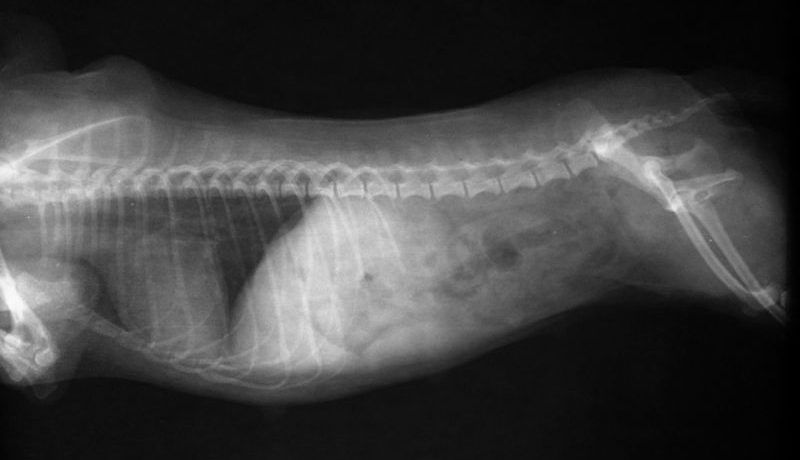
Radiography (X-rays) will allow your vet to visually examine your dog’s chest and abdomen to assess for evidence of heart disease, to check for abnormal accumulation of fluid and identify tumours.
Ultrasonography of the abdomen may be performed to measure the thickness of the intestines and look for evidence of enlarged lymph nodes, foreign bodies in the gastrointestinal tract or masses which could be cancer. Abdominal ultrasound can reveal changes to intestinal wall structure, including:
- Thickening without loss of normal layers (indicative of inflammatory bowel disease)
- Thickening with loss of layers (indicative of infiltrative intestinal neoplasia)
- ‘Tiger stripes’, a possible indicator of lymphangiectasia
Abdominal ultrasound will also identify an abnormal accumulation of fluid in the abdomen (ascites) or lungs (pleural effusion) and mesenteric lymphadenopathy, an enlargement of lymph nodes within the abdomen.
Endoscopy
A small camera, attached to a tube, is passed through the dog’s mouth and/or anus into the intestines so that the walls of the stomach and intestinal tract can be visually examined for ulcers, tissue masses (tumours) or abnormalities in the wall or cell structure.
During the endoscopic procedure, tissue samples can be taken from the stomach, intestines and rectum for biopsy. Analysis of biopsied tissue is very useful for determining why an animal is losing protein through its intestines and can provide a definitive diagnosis of lymphoma and secondary lymphangiectasia.
Expert advice from Dr Felicia:
Some patients with PLE may be too unwell/unstable for an anaesthetic to collect samples due to fluid accumulation in their chest/abdomen, or extremely low protein levels in their blood. Efforts may be made to initiate treatment based on available information and then samples taken later.
Surgical exploration
Open abdominal surgery or “keyhole” surgery of the abdomen (laparoscopy) may be performed to evaluate all the organs and collect multiple, full-thickness biopsies, which may be necessary to rule out certain types of disease like lymphangiectasia.
Prognosis
Even with aggressive treatment, sadly PLE can dramatically shorten the life expectancy of your dog, and untreated, it can be fatal. The sooner PLE is diagnosed, the better the long-term outcome. The condition varies in severity from mild to severe and prognosis depends on the underlying cause of PLE in each case. However, the long-term prognosis in most dogs is guarded.
As certain breeds show a predisposition to PLE or related diseases, including Yorkshire Terriers, Soft-coated Wheaten Terriers, Basenjis and Norwegian Lunehunds, dogs of these breeds showing any of the symptoms of PLE should immediately be examined. Soft-coated Wheaten terriers are known to have a median survival time of five months after diagnosis of PLE and of two months if they suffer from concurrent protein-losing nephropathy.
Prognosis may be worsened by the development of life-threatening complications such as pulmonary thromboembolism (a blockage in the arteries that supply blood to the lungs caused by blood clots) and decreased serum albumin concentration (hypoalbuminemia).

Treatment for protein losing enteropathy in dogs
As there are many underlying causes of PLE in dogs, there is no single treatment protocol for this syndrome and every patient has different needs. The therapeutic aims are to treat the underlying cause and to support the patient. In most cases, treatment will be determined by the underlying disease. Your vet will develop a treatment plan to help you manage your dog’s symptoms, which will include exercise and a special diet to facilitate the absorption of nutrients by your dog’s body.
Depending on the underlying causes of PLE identified by the diagnostic tests, treatment may include:
- Dietary changes such as hypoallergenic diet for IBD or an ultra-low fat, easily digestible diet for intestinal lymphangiectasia
- Anti-inflammatory medication is the main treatment for IBD but can have unwanted side effects when used at higher doses
- Procedures to remove the fluid from the chest or abdomen (e.g., thoracocentesis or abdominocentesis) which can provide some relief and stabilisation in the short-term
- Plasma transfusions can help to increase the protein level in the short-term though typically large volumes are required
- Intravenous fluids for patients who are not eating and are dehydrated
- Diuretics to reduce fluid build-up in patients with ascites or pleural effusion
- Antithrombotic therapy such as low dose aspirin to reduce the risk of forming blood clots
- Dietary supplementation with calcium if a low serum concentration of ionised calcium is detected
- Anti-emetics to treat nausea
- Tube feeding, in cases of severe vomiting or unwillingness to eat
- Synthetic colloids to restore plasma volume
- Vitamin B12 (cobalamin) injections or supplementation
- Deworming (for parasitic infections)
- Antiulcer medication (if gastric ulcers are present)
- Surgery
Management and long-term care of PLE in dogs:
PLE sufferers are fragile because of their hypoalbuminemia, gastrointestinal disease and their potential to develop spontaneous blood clots. Supportive care is therefore extremely important and providing adequate nutrition is vital.
Regular follow-up examinations and testing will be required, including complete blood counts and biochemical profiles to check that your dog’s blood protein level is stable and not becoming dangerously low. Your vet will also check your dog for symptoms such as breathing difficulties from fluid build up in the abdomen.
Expert advice from Dr Felicia:
Since PLE is a serious consequence of severe gastrointestinal disease, patients with PLE require close monitoring and may need a lot of supportive care/medications to help them to get well again, and treatments will likely be lifelong.
In conclusion
Any intestinal disease, if severe enough, can result in protein losing enteropathy (PLE) in dogs. The underlying cause of PLE is disruption of the intestinal lining, leading to the abnormal loss of protein from the intestines. Because several diseases can lead to this condition, PLE is classified as being a syndrome rather than a disease.
As there are a diverse range of underlying conditions that cause PLE, it may develop in dogs of any age, breed, or sex. However, some breeds have been demonstrated to be particularly at risk; these include Yorkshire Terriers, Soft Coated Wheaten Terriers, Norwegian Lundehunds, and Basenjis.
Many dogs with PLE have non-specific signs of gastrointestinal disease, however, the most common clinical signs are diarrhoea, vomiting, and weight loss. Diagnosis entails blood testing and other investigative tests to determine the underlying disease that may have caused the PLE. In general, treatment of PLE is determined by the underlying disease and the severity of the clinical signs. Modified nutrition is generally a part of the ongoing management of PLE. In some cases, the primary, underlying disease may not be treatable but even where aggressive treatment is undertaken, the long-term prognosis in most dogs with PLE is guarded.
Bow Wow Meow Pet Insurance can help protect you and your dog should an unexpected trip to the vet occur.
-
Find out more about our dog insurance options
-
Get an online pet insurance quote
Bow Wow Meow is proud to have been awarded winner of Canstar’s ‘Most Satisfied Customers’ Award in the Pet Insurance category for both 2024 and 2025!
Bow Wow Meow is proud to be the only Pet Insurer that has been awarded Product Review’s Top Rated Pet Insurance every year from 2018 to 2026! This is based on 3,193 independent customer reviews (as at 09/04/2026), with an overall rating of 4.5 stars.

We also have the following ratings on other review platforms:
Google Review rating of 4.5 stars (based on 1,358 reviews)
Trust Pilot rating of 4.6 stars (based on 555 reviews)
Bow Wow Meow has been chosen as Winner of 4 categories in the 2026 Finder Pet Insurance Customer Satisfaction Awards!


– Most Trusted Pet Insurance
– Easiest to Claim Pet Insurance
– Most Recommended Pet Insurance
– Legendary Service Pet Insurance
We also received Highly Commended Awards for Loved Brand Pet Insurance and Best Value Pet Insurance.
Bow Wow Meow is an Australian-owned and operated pet insurance provider. Our focus is purely on pets, and we are proud to have helped provide peace of mind to over 200,000 Aussie pet owners since 2008.