Eye (ocular) anomaly in dogs and cats
What is eye (ocular) anomaly in dogs and cats?
In medical terminology, an anomaly is an abnormality that is present at birth. An eye or ocular anomaly is a defect affecting the structure of the eye that is evident at birth, or shortly after. This is also known as a congenital disorder or birth defect.
The eyeball of dogs and cats is located within the bony socket of the head and is partially protected by the three eyelids.
The eyeball is a complicated organ comprised of several structures, including the following:
- Cornea – the transparent dome-shaped surface covering the front of the eye, it plays an important role in focusing vision.
- Iris – the coloured circle around the pupil, it changes the size of the pupil and allows varying amounts of light to enter the eye.
- Pupil – the opening in the centre of the iris that allows light to pass through to the retina at the back of the eye.
- Lens – a transparent structure behind the iris which ‘bends’ light rays to focus on the retina.
- Retina – the light-sensitive tissue that lines the inside of the eye and sends visual signals through optic nerve to the brain.
- Vitreous fluid – A clear, jelly-like fluid that fills the space between the lens and the retina.
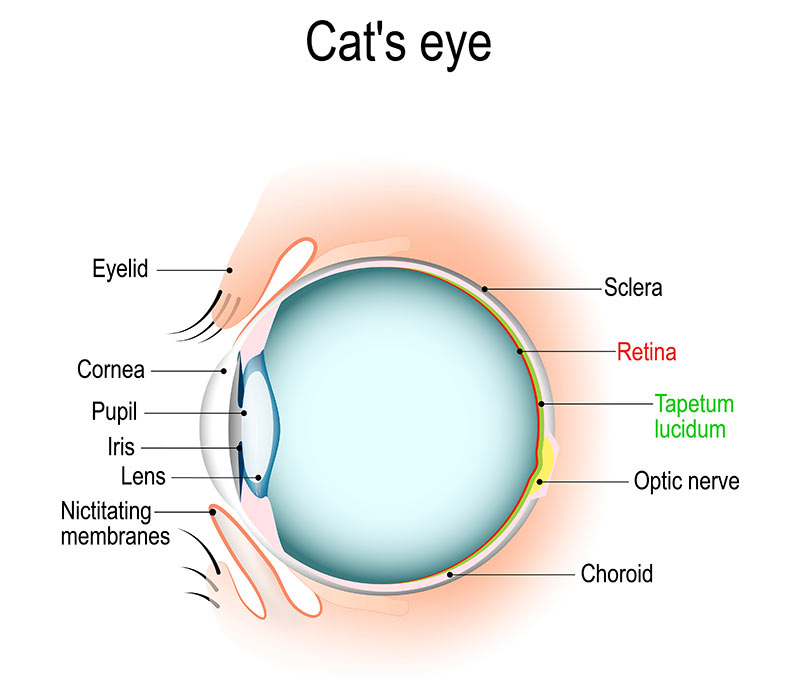
Cat feline eye anatomy diagram
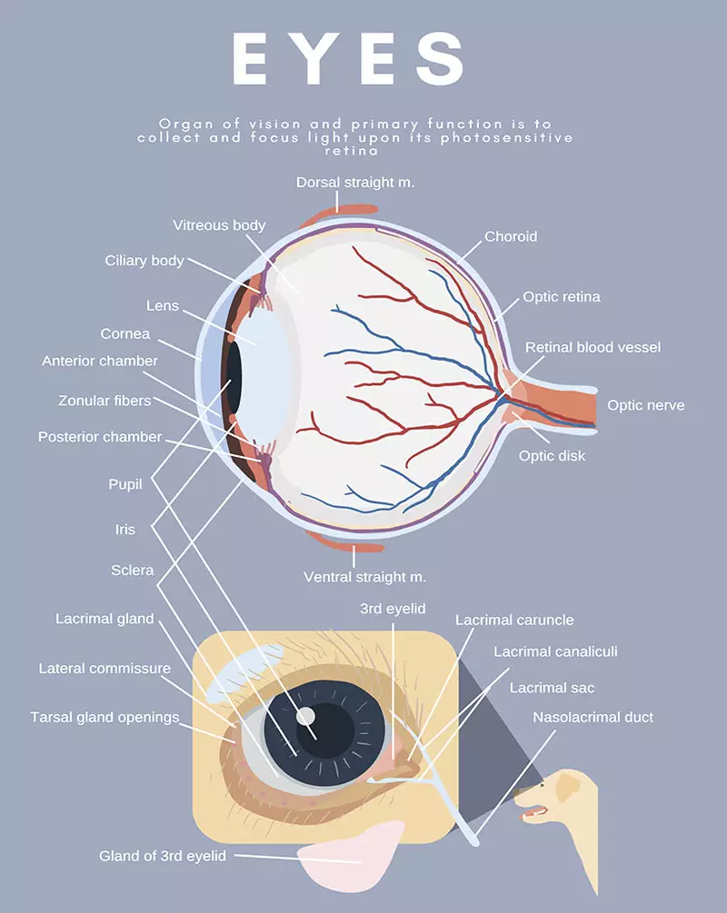
Dog canine eye anatomy diagram
The eyes convert reflected light into nerve impulses that are interpreted by the brain as images of the world. To do this effectively, all the various components of the eye need to be healthy. Unfortunately, many anomalies can disrupt the way the eyes function.
Congenital anomalies of the eyeball or its surrounding tissue can be evident in a dog or cat shortly after birth or may develop within the first six to eight weeks of life. Eye anomalies may be genetically inherited, can develop spontaneously or occur in utero. Exposure to toxic compounds, lack of nutrients, and systemic infections and inflammations during pregnancy are other possible causal factors for ocular abnormalities. Congenital and inherited ocular diseases are reported less frequently in the cat than the dog.
Because it is difficult to predict the costs of veterinary care, it can help to have measures in place to help prepare for the unexpected. Pet insurance can help by covering a portion of the eligible vet bill if the unexpected does happen.Find out more about our pet insurance options.
Eye anomalies that can occur in dogs and cats
Colobomas of the lid (Feline eyelid agenesis)
- From the Greek “koloboma”, meaning defect, eyelid coloboma is a congenital defect of the eyelid in which the upper lateral eyelid is typically not formed.
- It may appear as notch in the eyelid, or tissue of the eyelid may be missing; it usually affects the upper lid and is often bilateral.
- Eyelid agenesis occurs sporadically in domestic cats and is rare in dogs.
- Appears to be inherited in Burmese, Persian, and Siamese cats.
Colobomas of the iris
- Misshapen or thin iris (coloured part of the eye).
- Does not typically affect vision and does not progress to anything else.
- Relatively uncommon in dogs and cats.
- Most are genetic; most commonly seen in Australian Shepherds and other herding dogs.
Congenital cataracts
- These are cataracts (opacity if the lens of the eye) that are present at birth.
- They usually occur in both eyes.
- Infections or toxins may cause the formation of these cataracts while the puppies are still in utero.
- Often inherited (e.g., Cavalier King Charles Spaniels, Miniature Schnauzers)
- Dogs suffer from cataracts more commonly than any other species.
Collie eye anomaly (Scleral dctasia syndrome)
- An inherited congenital condition resulting from a mutation if the chromosomes that determine the development of the eyes (it always occurs in both eyes).
- This anomaly impairs vision and can result in other eye abnormalities such as a detached retina, optic nerve abnormalities and a loss of retinal cells.
- Originally discovered in the Collie breed, collie eye anomaly also affects other breeds including Australian Sheepdogs, Border Collies and Shetland Sheepdogs (‘Sheltie eye’).
- Approximately 70 to 97 percent of rough and smooth collies in the United States and Great Britain are affected.
Collie Eye Anomaly (Scleral Ectasia Syndrome) | Source
Congenital keratoconjunctivitis sicca (KCS)
- Also referred as dry eye, KCS is a failure of tear production that leads to painful damage to the surface of the eyes.
- Common in cats, but in dogs unique to the Cavalier King Charles Spaniel, this disease is often accompanied by ichthyosiform dermatitis which is caused by abnormal rates of cell turnover in the skin; both conditions are present from birth in affected puppies.
Corneal sequestrum
- Appearing as a dark brown or black spot in the cat’s eye, a corneal sequestrum is a piece of dead corneal tissue.
- The likelihood of total vision loss – or, in severe cases, loss of the eye – is increased when the sequestrum is large and extends deep within corneal tissue.
- Brachycephalic breeds, characterised by their broad skull shapes, have a higher chance of developing corneal sequestrum.
Dermoid cysts
- Tumour-like cysts present at birth on the eyelid, conjunctiva or cornea.
- They are non-cancerous and usually harmless.
- They are fairly common in domestic dogs and rare in domestic cats.
- A hereditary component is strongly suspected in dogs.
Entropion
- Entropion is a congenital condition in which the eyelids appear to roll inward against the cornea of the eye; this can cause a great deal of discomfort for the animal.
- It is more common in dogs then cats.
- Breeds that are commonly seen with entropion include but are not limited to; Chow Chow, Chinese Shar-Pei, Irish Setter, Golden Retriever, Labrador Retriever, Collie, Great Dane, and Rottweiler.
Lens luxation
- The support ligaments of the lens weaken or break causing the lens to dislocate from its normal position.
- Weakness of the lens ligaments is known to be hereditary in the terrier breeds, Chinese Shar Pei and Border Collie.
Microphthalmia
- The eyeball is smaller than normal and usually the internal structures of the eyeball are also abnormal, usually resulting in visual impairment or completely blindness.
- Microphthalmia is inherited in Australian Shepherds, Great Danes, Beagles, Collies, Borzoi, Dobermans, Sealyham Terriers, Bedlington Terriers, Portuguese Water Dogs, Shelties, Akitas, Miniature Schnauzers, Labrador Retrievers, Dachshunds and Cavalier King Cocker Spaniels.
- It can also occur in new-borns whose mothers received certain medications during pregnancy.
Persistent hyperplastic tunica vasculosa lentis (PHTVL) / persistent hyperplastic primary vitreous (PHPV)
- Begins in utero, with progressive atrophy of the vascular system (blood vessels) that supports the lens of the eye, often leading to cataracts.
- Appears to affect certain breeds more than others with Doberman Pinschers being among those most affected by the condition; also found in Staffordshire Bull Terriers, Basenji, Briards, Cocker Spaniels, Beagles and Rottweilers.
Persistent pupillary membrane
- A persistent pupillary membrane occurs when strands of foetal tissue remain within the eye chambers after birth; these fibrous strands may actually crisscross the eye chambers and attach to the lens and/or cornea.
- A corneal opacity may be seen.
- The condition may be inherited in Basenjis, Pembroke and Cardigan Welsh Corgis, Chow Chows and Mastiffs.
Photoreceptor dysplasia
- A malformation of the cells that perceive light and colour, indicated by slow or absent pupillary reflex to light (when pupil does not contract or dilate normally).
- May result in involuntary eye movement and poor ability to see in both low light and daylight.
- In cats, breeds more prone include Abyssinian, Persian and Domestic Shorthair cats.
- In dogs, breeds more prone include Collies,Irish Setters, Miniature Schnauzers and Norwegian Elkhounds.
Primary congenital glaucoma (PCG)
- PCG is defined as high pressure within the eye without any other ocular cause, it is a serious condition which causes irreversible damage of the optic nerve.
- An inherited eye disorder affecting cats, although PCG can also affect dogs.
- Burmese, Siamese and Persian cats are among the predisposed breeds.
Progressive retinal atrophy and degeneration (PRA /PRD)
- Progressive retinal atrophy (PRA) includes several inherited diseases that lead to degeneration of retinal photoreceptor cells (rods and cones) in cats and dogs, typically leading to blindness.
- Some breeds experience an earlier onset than others; other breeds do not develop PRA until later in life.
- PRA worsens over time, with affected animals typically experiencing night blindness initially because the rods are affected first. The condition progresses to failed daytime vision.
- Feline Progressive Retinal Atrophy (PRA-rdAc), caused by a mutation in a specific gene, has been documented in 37% cat breeds, with high frequency in North American and European Siamese populations.
Retinal detachment
- Retina detaches from the back of the eye causing blindness
- Common in Labrador Retrievers, Bedlingtons, and Sealyham terriers
Retinal dysplasia
- Retinal dysplasia refers to a malformation of the retina in which the cells and layer of retinal tissue do not form properly during embryonic development.
- Usually first noticeable in puppies about six weeks of age; instead of a flat appearance, the retina appears to be layers of folded tissue (this can only be seen with the aid of an ophthalmoscope).
- Retinal dysplasia generally results in inadequate vision and can lead to blindness.
- It is considered an inherited trait and various forms of retinal dysplasia are found in many dog breeds, including English Springer Spaniels, Briards, Labrador Retrievers, Sealyham Terriers, Bedlington Terriers, Doberman Pinschers, Cocker Spaniels, Rottweilers, Yorkies and Old English Sheepdogs.
- It also affects kittens that have been exposed and infected with feline leukaemia or feline panleukopenia, either while in utero or after birth.
Symptoms of eye (ocular) anomaly in dogs and cats
Symptoms of eye anomalies vary considerably and depend largely on the abnormality that is present. In some cases, there may be no observable symptoms (the eye may continue to look normal), and the anomaly can only be identified by ophthalmic examination.
Signs of visual impairment may be difficult to detect because vision may be gradually affected, and animals are remarkably adaptable to progressive blindness and can often seem to perform normally in their familiar environments. Evidence of the visual impairment is more pronounced if the furniture is rearranged or if the animal is in unfamiliar surroundings. Night blindness may be evident by reluctance to go down stairs or down a dark hallway.
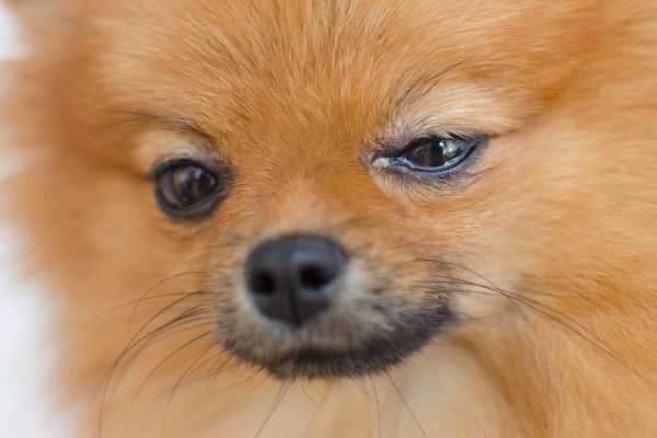
Physical signs of an eye anomaly may include:
- Redness
- Excess tearing, watery eyes
- Squinting
- Sensitivity to bright light
- Eyelid twitching
- Involuntary eye movements
- A bulging or enlarged eyeball or iris
- An over-dilated or under-dilated pupil
- An obvious physical deformity to the eyeball, such as an abnormally small eyeball, missing eyeball or hidden eyeball
- A misshapen eyelid, pupil or iris
- A lack of iris or pupil
- A growth or protrusion on the eye or eyelid
- A white, cloudy appearance to the eye
Causes of eye (ocular) anomaly in dogs and cats
Congenital anomalies of the eye may be the result of one or more genetic, infectious, nutritional or environmental factors; it is often difficult to identify the exact cause or causes. Retinal dysplasia, for example, is a maldevelopment of the retina that may arise from trauma, genetic defect or intrauterine damage such as a viral infection.
Causes of ocular anomalies in dogs and cats may be broadly grouped as:
- Genetic
- Developmental malformation
- Deformation
- Disruption
- Environmental
Genetic causes:
- Many eye conditions are recessively inherited.An animal may be carrier of the defective gene and not itself develop the disorder, but when two carriers mate, their offspring will have a 25% chance of being affected, a 50% chance of being a asymptomatic carrier, and a 25% chance of being unaffected and not a carrier.
- In cats, the Persian, Burmese, and Siamese are among the breeds repeatedly linked with eye anomalies of genetic origin.
- Most forms of retinal dysplasia in dogs are inherited, and many DNA mutations have been reported.
- Progressive retinal atrophy (PRA) has been shown to have a genetic component, and many breeds of dogs and cats are suspected of having inherited PRA.
- Feline Progressive Retinal Atrophy (PRA-rdAc) is caused by a mutation in CEP290 gene, and has been documented in 37% cat breeds, with high frequency in North American and European Siamese populations.
- The cause of collie eye anomaly is a defect in chromosome 37, occurring in dogs with parents that the carry the genetic mutation. It is suspected that other genes may also be involved.
Developmental malformation:
- The anomaly is developmental in origin, meaning that it develops in utero while the animal’s cells, tissues and organs are being formed; there is abnormal embryonic or foetal development.
- Problems in the developmental process can lead to congenital eye anomalies such as anophthalmia (no eye), microphthalmia (small eye), coloboma (failure of the optic fissure to close), aniridia (absent or partial iris), and optic nerve hypoplasia (underdeveloped optic nerve).
- A dysplasia is an abnormality in form or development, for example, retinal dysplasia can result from abnormal formation of the retina during embryonic development.
Deformation:
- There is a change from the normal size or shape of an anatomic structure due to mechanical forces that distort an otherwise normal structure.
- Deformations occur most often late in pregnancy or during delivery.
Disruption:
- The events that occur when a foetus that is developing normally is subjected to a destructive agent such as infection or inflammation.
- Maternal viral infections, especially during early foetal development, can result in multiple ocular anomalies in cats (e.g., panleukopenia virus) and dogs (e.g., herpesvirus).
Environmental causes:
- An agent that can cause a birth defect is known as a “teratogen”.
- A drug, toxin or a disease affecting a pregnant mother can increase the chance for the baby to be born with a birth defect.
- Nutritional deficiencies during pregnancy can be a contributing cause to birth defects.
How is eye (ocular) anomaly in dogs and cats diagnosed?
Medical history:
A complete medical history of the dog or cat will be required, including any relevant in utero conditions such as exposure to toxins, and the animal’s development and environment after birth.
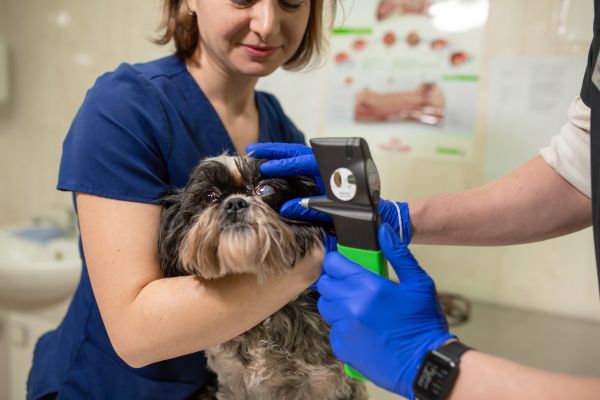
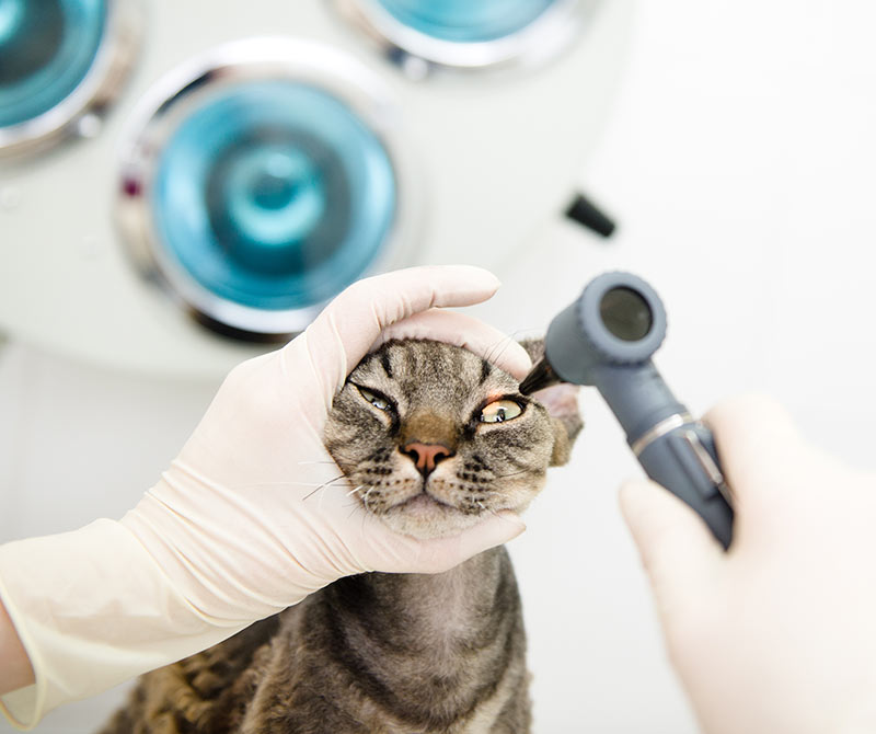
Ophthalmic examination:
Characteristic changes in the retina and other parts of the eye may be observed through an ophthalmic examination by the vet or veterinary ophthalmologist. These tests are usually painless and the animal does not have to be anesthetised.
- Abnormalities within the eye will be examined with an ophthalmoscope and/or a slit-lamp biomicroscope.
- More sophisticated tests such as electroretinography may also be used.
- A Schirmer tear test may be used to see if the eyes are producing an adequate amount of tears.
- If high pressure in the eye (glaucoma) is suspected, a diagnostic tool called a tonometer will be applied to the eye to measure its internal pressure.
- Gonioscopy, an examination of the part of the eye between the cornea and the iris, may be used for detection of glaucoma.
Ultrasound:
An ultrasound of the eyes may detect anomalies associated with the lens, the vitreous humour (the clear fluid which fills the space between the lens and retina), the retina, or other problems that are taking place in the posterior (back) segment of the eye.
Angiography:
A diagnostic method that can be used for viewing problems in the posterior of the eye, such as detachment of the retina and abnormal blood vessels in the eye. An angiogram uses x-rays and a special dye that is visible on X-ray, which is injected into the area that needs to be visualized, so that the blood vessels can be examined for irregularities.
Prognosis
 Although eye anomalies are not fatal, more severely affected animals may be partially or totally blind. Many of the conditions have no cure and no accepted treatment for slowing the progression or prolonging vision. If vision is only mildly affected by the condition, the animal can lead a full and happy life. However, even blind dogs may continue to do well in familiar surroundings by relying on their other acute senses.
As most congenital ocular anomalies are hereditary, animals that have been diagnosed with any of these disorders should not be bred. Breeders should always have their stud animals screened for any eye abnormalities because this is the only way of reducing the risks of their offspring being born with congenital eye disorders. Therefore, when buying a breed that is known to be prone to developing a congenital eye anomaly, it is important to deal with responsible breeders who routinely have their animals’ eye tested.
Treatment for eye (ocular) anomaly in dogs and cats
Treatment will depend on the specific type of eye abnormality that is affecting the dog or cat; however, for many eye anomalies there is no treatment that can help. Surgery can repair some congenital birth defects, and medicines can be used to mitigate the effects of some types of defects. There is currently no treatment available for many eye anomalies, including retinal dysplasia, progressive retinal atrophy, microphthalmia and persistent pupillary membrane.
Surgical treatment:
- Cataracts: The only current treatment option is surgery but this may not be necessary unless a high degree of vision is lost; in this case, sight can be restored to near normal.
- Colobomas: Numerous surgical procedures exist, depending on the size of the defect and the impact it is having on the eye.
- Entropion: Treatment is always surgical repair.
- Dermoid cysts: Usually a simple surgical procedure, depending on the size and location of the cysts.The dermoid mass is dissected from the normal eye tissue; those located on the cornea present the greatest challenge.
- Detached retina: Surgery may be effective in preventing or in re-attaching a detached retina.
- Laser surgery and cryosurgery, which uses extreme cold to destroy unwanted cells or tissue, may be used in some cases.
Medical treatment:
- Congenital keratoconjunctivitis sicca (KCS), commonly known as dry eye, can often be treated with tear substitutes in combination with antibiotics.
- Mydriatics, a type of drug that dilate the pupil, may be used to increase vision when congenital cataracts are present.
- Artificial tears for temporary relief from dryness.
- Topical antibiotics to treat or prevent bacterial infections.
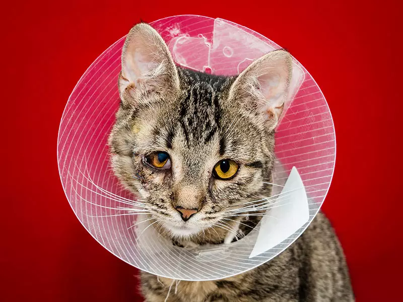
Ongoing treatment and management:
- Extra care and lifestyle adaptations are needed for animals with vision impairment, in order to help them in living comfortably.
- Regular consultations with the vet or specialist may be necessary for ongoing assessment of the condition.
- In cases of photoreceptor dysplasia, there is no medical treatment that will delay or prevent its progress, but dogs with this condition generally do not suffer from any other physical abnormality and can learn to manage their environment very well, as long as they are able to depend on their environment being stable and safe.
- Congenital KCS requires frequent check-ups with a veterinarian to monitor tear production and the status of the external eye structures.
- Abnormalities such as congenital cataracts, PHTVL, and PHPV require checkups twice yearly to monitor progression.
- Dog goggles that function as sunglasses can be used to reduce glare and subsequent squinting where there is sensitivity to light.
- Since most congenital ocular anomalies are hereditary, you should not breed a dog that has been diagnosed with any of these disorders.
In a nutshell
 The eye is a complicated organ and there are a variety of abnormalities that can affect an animal’s eye or surrounding tissues. Congenital abnormalities of the eyeball or its surrounding tissue are generally evident shortly after a puppy’s birth or may develop within the first six to eight weeks of life. Most eye anomalies are genetically inherited, but some develop spontaneously. Exposure to toxic compounds, lack of nutrients, and systemic infections and inflammations during pregnancy are other potential risk factors for ocular abnormalities.
In cats, several defects are common in breeds such as the Persian, Himalayan and Burmese cats that affect the anterior, or front section, of the eye. Examples are colobomas (agenesis of the eyelids), dermoid cysts and corneal sequestrum. In dogs, other problems such as cataracts, lens luxation and retinal dysplasia, cause problems of the intraocular structures (those within the eyeball), are more common.
Treatment will depend on the particular anomaly that is present. Surgery or medical management may be recommended; however, many conditions currently have no available treatment. If vision is only mildly affected by the condition, the affected animal can go on to lead full and happy lives. However, if sight is more seriously impacted by the condition, then owners will need to take extra care when looking after their pets.
Bow Wow Meow Pet Insurance can help protect you and your pet should an unexpected trip to your vet occur.
- Find out more about our dog insurance options
- Find out more about our cat insurance options
- Get an instant online pet insurance quote
Bow Wow Meow is proud to have been awarded winner of Canstar’s ‘Most Satisfied Customers’ Award in the Pet Insurance category for both 2024 and 2025!
Bow Wow Meow is proud to be the only Pet Insurer that has been awarded Product Review’s Top Rated Pet Insurance every year from 2018 to 2026! This is based on 3,193 independent customer reviews (as at 09/04/2026), with an overall rating of 4.5 stars.

We also have the following ratings on other review platforms:
Google Review rating of 4.5 stars (based on 1,358 reviews)
Trust Pilot rating of 4.6 stars (based on 555 reviews)
Bow Wow Meow has been chosen as Winner of 4 categories in the 2026 Finder Pet Insurance Customer Satisfaction Awards!


– Most Trusted Pet Insurance
– Easiest to Claim Pet Insurance
– Most Recommended Pet Insurance
– Legendary Service Pet Insurance
We also received Highly Commended Awards for Loved Brand Pet Insurance and Best Value Pet Insurance.
Bow Wow Meow is an Australian-owned and operated pet insurance provider. Our focus is purely on pets, and we are proud to have helped provide peace of mind to over 200,000 Aussie pet owners since 2008.