Skin and coat conditions in dogs: signs, causes and treatment


Summary
Skin and coat problems are common in dogs and can be caused by allergies, parasites, bacterial or fungal infections, inherited disorders, hormonal or metabolic disease, nutritional imbalances and skin tumours. Common signs include itching, excessive licking or scratching, redness, sores, scaling, unusual odour, hair loss and changes in the texture or appearance of the coat. Many skin conditions respond well to appropriate treatment, but some are recurrent or require lifelong management. Because different conditions can cause very similar symptoms, a veterinary examination is often needed to identify and treat the underlying cause.
Continue reading below to learn more about dog skin and hair coat conditions including:
- An A to Z of many of the more common conditions
- Symptoms of hair coat and skin conditions in dogs
- Causes of hair coat and skin conditions in dogs
- Diagnosis of hair coat and skin conditions in dogs
- Treatment for hair coat and skin conditions in dogs
Why are healthy skin and coat important?
The skin is the largest organ of a dog’s body, and most of it is protected by the hair coat. Together, the skin and coat form an important barrier against physical injury, ultraviolet light, infectious organisms, irritants and excessive water loss. They also contribute to sensation and body-temperature regulation.
Healthy skin should generally be supple and free from persistent redness, sores, scaling, excessive oiliness or unpleasant odour. A healthy coat is typically smooth and appropriate for the dog’s breed and coat type, without unexplained thinning, bald patches or excessive shedding.
Changes in the skin or coat may be caused by a local problem, such as parasites, infection or irritation. They can also be an early sign of an underlying allergy, hormonal disorder, nutritional imbalance or other health condition. Persistent itching, hair loss, lumps, sores or changes in coat quality should therefore be discussed with a veterinarian.
Because the size, shape, and length of hair are controlled by genetics, different breeds have different coat characteristics:
- Breeds such as smooth-haired terriers and toy poodles have numerous follicles that produce plentiful guard hairs and fewer under-hairs.
- Breeds such as German Shepherds, Airedales and Rottweilers have only half as many follicles but have twice as many under-hairs sprouting from each follicle.
- Other breeds such as Siberian Huskies, Alaskan Malamutes and many of the Retriever breeds have long, thick hair coats with both an outer coat of guard hairs and an undercoat of fine hair that serves as an insulating layer.
- Many short-haired breeds lack a distinctive undercoat and may shed hair in low levels all year round.
Common skin and coat conditions in dogs: A–Z
Acute moist dermatitis, pyotraumatic dermatitis or “hot spots”
Hot spots, also known as acute moist or pyotraumatic dermatitis, are rapidly developing areas of inflamed and self-traumatised skin. Persistent licking, chewing or scratching damages the skin, and bacterial overgrowth or infection may develop within the affected area.
It is typically a self-inflicted trauma resulting from the dog’s attempts to alleviate pain or pruritus (intense itching). Licking, biting and scratching the area by the dog irritates the inflamed skin even more. The condition is also known as ‘pyotraumatic dermatitis’ because the self-trauma is a major factor in the development of the lesions or hot spots.
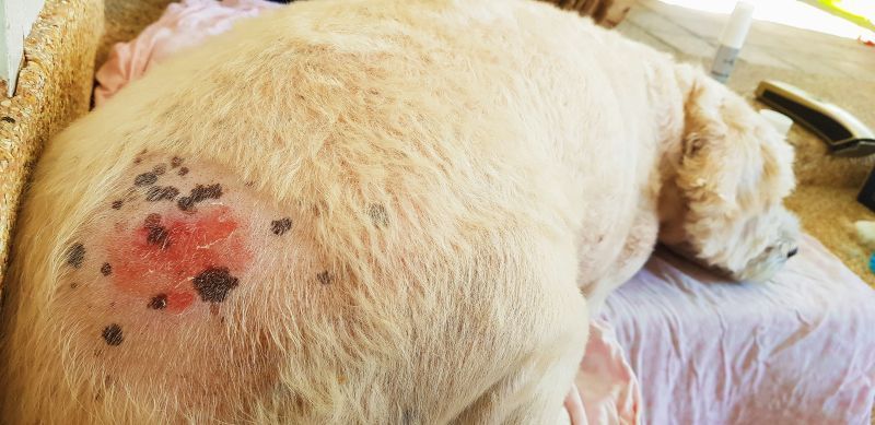
Hot spots are typically circular lesions that are moist, raw, inflamed and hairless. They are usually found on the head, near the tail base and other areas on the trunk, and can change dramatically in size in a very brief period.
Occurring in both inside and outside dogs, hot spots are more prevalent in hot and humid weather and less frequent in the colder temperatures. Many dogs develop several of these lesions over the course of their lives. However, this is not a long-term condition; a lesion may suddenly appear, be treated and disappear in less than a week. Another lesion may suddenly appear a short time later, the following year or may never occur again.
Common underlying causes include a bacterial infection following a minor skin injury, parasites such as mites and fleas, a local allergic reaction to a specific antigen, ear infections, poor grooming, burs or plant awns and anal gland disease.
Expert advice from Dr Felicia:
Hotspots tend to be localised to one area, and usually in dogs with thick, long-haired coats, where moisture more easily gets trapped and allows the skin infection to spread and become severe quite quickly. Thankfully they also tend to resolve well and swiftly with treatment.
Abnormal skin growth syndromes (hyperplastic and seborrheic syndromes)
These are a group of conditions involving abnormal growth of the skin cells, leading to scaling and thickening of the outer layer of skin. Abnormal skin growth syndromes include ichthyosis, familial footpad hyperkeratosis, granulomatous sebaceous adenitis, hereditary congenital follicular parakeratosis and psoriasiform-lichenoid dermatosis (for more information on some of these conditions, see below).
Acanthosis nigricans
Acanthosis nigricans is a disorder of pigmentation in which there is abnormal darkening, or hyperpigmentation, of the skin.
Primary acanthosis nigricans seems to exclusively affect Dachshunds less than two years of age. The skin darkens and thickens, seborrhoea (see below) develops, and secondary infections with bacteria or yeast can occur. Large areas of the body may be affected. This condition is rare and is diagnosed mainly through breed history and biopsy.
Secondary acanthosis nigricans has several causes and can occur in any breed of dog and at any age. It is mainly caused by obesity, endocrine disease or allergies. In this form, the pigment melanin mobilises to areas of chronic inflammation, resulting in the dark appearance of the skin. Hair loss and itching are likely to occur. The disorder may also develop as a secondary stage of another skin disease.
Acariasis
see Mite infestation (mange, acariasis, scabies)

Acquired aurotrichia
This hereditary condition affects Miniature Schnauzers. In affected dogs, the primary hairs along the middle of the back above the spine changes from the normal colour to golden. Additionally, the secondary hairs thin out, giving the coat a rough feel to the touch. Acquired aurotrichia usually starts in young adulthood, at around two and half years. Typically, there are no other changes to the skin and no related health problems. In most dogs, the coat colour returns to normal within two years.
Albinism
Albinism is a rare condition in dogs that is different from extreme white spotting (white areas on the coat). True albinism is a genetic condition in which pigmentation of the eyes, coat and skin is completely absent. Albinism is always accompanied by pale irises (i.e., the coloured part of the eyes is usually pink) and vision defects. Affected animals are at increased risk of skin cancer from sun exposure, especially in areas of skin with short or thin hair.
Alopecia
Alopecia, or hair loss, is a dog skin condition that can have numerous underlying causes. It can be hereditary, congenital, or acquired.
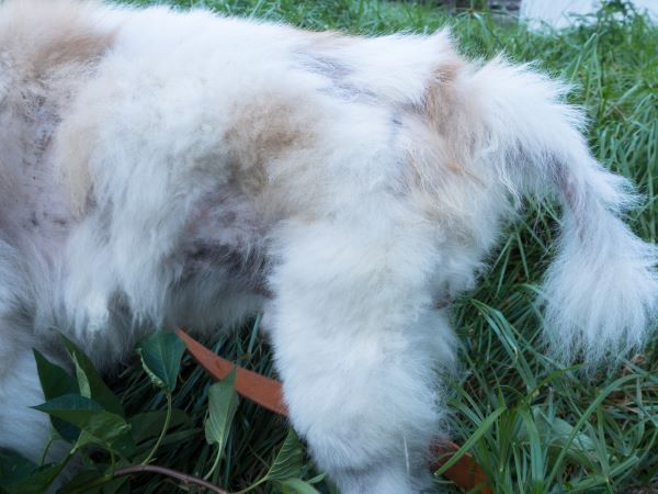
Acquired alopecia often results from intensive scratching or licking of an itchy area as the result of an underlying skin condition such as ringworm, yeast or bacterial infection, or common skin parasites such as fleas, ticks, mites or lice. This is referred to as‘inflammatory acquired alopecia’, the most common form of the condition.
Seasonal flank alopecia can appear in Boxers and Airedale Terriers. Various woolly syndromes and post-clipping alopecia can occur in Spitz-type breeds. Another condition called alopecia X occurs in Pomeranians and other breeds.
In cases of hereditary alopecia, dogs may be born either totally or partially without hair. Hairlessness can also develop later in life in dogs that are born with normal coats, with alopecia occurring when the dog sheds its juvenile coat or becomes a young adult.
Colour dilution alopecia is a form of hereditary alopecia typically causing hair loss in dogs with blue (dilute black) or fawn (dilute brown) coat colours. Most affected dogs are born with normal hair coats, but during their first year they begin to develop hair follicle inflammation, brittle hair and progressive, patchy hair loss in the blue or fawn-coloured areas of the coat, resulting in a ‘moth-eaten’ appearance (other areas remain normal).
Black hair follicle dysplasia is a type of hereditary alopecia resulting in hair loss, localised to black or dark hairs of dogs. Dogs are usually affected at a young age, and are usually bicolour or tricolour. The condition is most common in Papillons and Bearded Collies.
Expert advice from Dr Felicia:
Hereditary alopecia is uncommon in dogs, but tends to be more evident at a young age. In contrast, older dogs with alopecia in the absence of other skin disease are more likely to be affected by an underlying systemic, endocrine cause.
Bacterial skin infections (also see pyoderma)
Species of bacteria normally reside on dogs’ skin without causing any problems. However, when the skin is more vulnerable than usual from dryness, inflammation or a wound, there is an increased risk of bacteria penetrating the skin and infection developing.
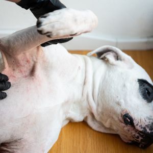
The most common species of bacteria associated with skin infections in dogs is staphylococcus. Signs of bacterial infection are often found in the skin folds, or around areas of broken and irritated skin. These may be red, itchy patches of pustules that seep blood and pus when they break. Severe infections may develop into painful ulcerations and boils.
Bacteria flourish in moist environments, so many dog skin infections are secondary to another condition which adds oil and moisture to the skin, such as a flea, tick or yeast infestation, an allergic reaction, or an underlying disease that depletes the immune system.
Expert advice from Dr Felicia:
Like humans, dogs can get resistant skin bacterial infections. Dogs that are on frequent courses of antibiotics are more likely to have resistant bacterial infections. Thankfully most resistant skin infections are still susceptible to topical antiseptics and antimicrobial shampoos.
Basal cell tumours
Basal cells lie at the base of the epidermis (the top layer of the skin). Basal cell tumours are common in middle-aged to older dogs, and are typically benign, though malignant tumours can occur.
Basal cell tumours develop on the outer layer of skin and appear as firm, elevated, hairless masses, usually around the head, neck, chest and back. The lumps, which are often hairless or ulcerated can vary in size are sometimes pigmented. Cysts may also form.
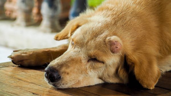
Although benign, basal cell tumours can cause ulceration and inflammation. Surgical removal is an effective treatment and reduces the chance of secondary infection and inflammation. Many breeds are predisposed to basal cell tumours, especially American and English Cocker Spaniels, Kerry Blue and Wheaten Terriers.
Canine ichthyosiform dermatosis
see Ichthyosis
Collagenous nevi
Occurring singularly or in multiples in dogs, these are benign (non-cancerous) nodules or collections of fibrous tissue made up of bundles of collagen. This is a rare condition in dogs.
Collie nose (Nasal solar dermatitis)
‘Collie nose’ is a condition in which breeds with little or no pigment on their faces develop lesions, usually on the nose, eyelids and adjacent areas, and occasionally on the haired bridge of the muzzle. The lesions appear as pink, raw areas around the nose and on the eyelids which may ulcerate and develop a crusty scab-like covering. This disease can progress to a more invasive cancerous form called squamous cell carcinoma.
Collie nose is thought to be caused by a hypersensitivity to solar radiation or sunlight (hence the term “nasal solar dermatitis”). It is more prevalent in areas with sunny climates, and lesions are worse in the summer months. Exposure to sunlight should be restricted in affected animals.
Expert advice from Dr Felicia:
Reducing access to sunlight is helpful for these patients, and may involve only going outdoors very early or late in the day to reduce UV exposure. There are also some products that help to ‘shield’ or protect the vulnerable areas.
Cutaneous asthenia (also known as dermatosparaxis or Ehlers–Danlos syndrome)
This is a condition in which the skin does not produce enough collagen (a protein that gives structure to the skin), resulting in loose, stretchy, fragile skin that hangs from the body, and associated joint and eye problems. It is seen in the Chihuahua, Dobermann, Labrador and Toy Poodle.
The fragile skin is evident from birth, while older animals develop hanging folds of skin and exhibit extensive scarring. Wounds heal slowly or not completely, and cysts and bruising may develop. The disease is not fatal in dogs.
Cutaneous mucinosis
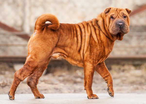
In this familial problem in some lines of Chinese Shar Peis, there is an abnormal accumulation of a protein called mucin in the skin, causing pronounced folding and blisters of the skin in some young dogs. As these dogs mature, the syndrome often becomes less severe, with many dogs seeming to improve as they get older.
Cutaneous papillomas (warts)
Warts are generally caused by papillomaviruses that are thought to be transmitted by direct contact with infected animals or indirect contact with contaminated objects, such as bedding. They usually occur on the dog’s face including the lips, tongue and inside of the mouth.
A wart is typically a hardened, light-coloured bump with a cauliflower-like appearance. Multiple warts are more commonly seen in younger dogs, and single warts are more common in older animals. Warts caused by viruses are usually benign and will generally disappear on their own within several weeks or months.
Expert advice from Dr Felicia:
Commonly seen in young dogs and frequent socialisers at the dog park. Thankfully most are self-limiting, though in patients with large numbers of warts that are painful in the mouth, cautery or cryotherapy can be performed.
Dermatitis or inflammatory disease
Dermatitis is a general term for skin inflammation of any type, rather than a specific diagnosis. It typically manifests as red, scaling, thickened skin characterised by itching, hair loss, abnormal smell and/or oiliness. It may progress to crusting, scaling, boils, blisters and other consequences of long-term inflammation; secondary bacterial and yeast infections often develop.
Possible causes of dermatitis include external agents acting as irritants or allergens to the dog’s skin, infections, burns, trauma or a systemic disease. The most common types of dermatitis in dogs are due to parasites, allergies and infections.
For more information, see Dermatitis in dogs and cats.
Expert advice from Dr Felicia:
Most dogs with skin disease will have an inflammatory component especially if there is any sort of infection involved. Chronic inflammation can result in thickening and changes in pigmentation of the skin, known as ‘lichenification’.
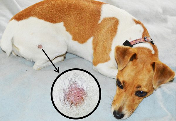
Dermatophytosis (ringworm)
This is the medical term for the fungal infection affecting the top layers of skin and hair that is more commonly known as ringworm. The organisms that are responsible are not worms but the fungi Microsporum canis, Trichophyton mentagrophytes, and Microsporum gypseum.
Ringworm is usually transmitted by direct contact with an infected animal, and appears as circular areas of hair loss throughout the body. It is diagnosed more commonly in young dogs than in adults.
Dermatosparaxis
Dermoid cysts
Usually found on skin above the backbone, dermoid cysts are skin pockets into which dander, hair, oil, and other debris accumulate. They are usually a congenital lesion, but not obvious until almost one year of age. While they can be solitary or multiple, most dermoid cysts are multiple and contain fully formed hair shafts. Although they are benign, they can be surgically removed if desired.
Ehlers–Danlos syndrome
Elbow calluses
A thickening of the skin at the elbow, calluses frequently occur in giant, short-haired breeds of dogs that sleep on hard surfaces such as wood or concrete. The callus forms to protect the bony elbow from pressure or trauma whenever the dog lies down. They can become infected and may bleed.
Expert advice from Dr Felicia:
These can also occur on the hocks of some dogs who like to lie down on hard ground. Usually the elbow lesions are more pronounced.
Epidermolysis bullosa (EB)
This is a rare, hereditary, congenital defect of the collagen gene that affects the attachments between the outer and inner layers of the skin, resulting in blistering of the skin. In affected dogs, blisters may be present at birth or develop within the first weeks of life. Minor skin trauma results in separation of the skin layers. Blisters form and soon rupture, leaving painful erosive lesions. The disease is usually fatal, with many affected puppies not living past 3 months of age.
Exfoliative dermatoses
Exfoliative dermatoses are a group of inflammatory skin disorders that are characterised by the excessive shedding of skin cells (flaking, scaling skin or dandruff); the term “exfoliative” refers to exfoliation, or shedding. There are many underlying causes of these conditions, from stress to nutritional deficiencies to allergies.

Familial footpad hyperkeratosis, hereditary footpad hyperkeratosis (HFH)
This is an inherited condition in which there is increased thickness of the hard, outer layer of the skin of the paw pads. All footpads are affected from an early age, although the disease is not usually congenital (present at birth). When hyperkeratosis is severe, horns, cracks (fissures) and secondary infections occur, causing pain and lameness. No other skin abnormalities occur.
Hyperkeratosis can also occur secondary to canine distemper, zinc responsive dermatosis or pemphigus foliaceus, which is an immune mediated disease.
Fibromas
Fibromas are isolated, generally raised, often hairless lumps originating on or under the skin surface, usually appearing on the head and legs. While these tumours are benign, surgical removal is recommended if they change appearance or grow large.
Granulomatous sebaceous adenitis
This hereditary skin condition destroys the dog’s oil glands in young or middle age. It is associated with a scaling skin and hair loss. First appearing when the dog is a young adult, it occurs in Standard Poodles and Akitas. A form of sebaceous adenitis has been reported with other conditions including hypothyroidism.
Hamartomas
see Skin tags
Haemangioma
Haemangiomas are small masses of blood vessels that appear as single or multiple, circular, often compressible, red or black lumps, resembling a “blood blister.” It is in fact a benign vascular (blood vessel) tumour of the skin in adult dogs, originating either in the dermis or the subcutaneous layer of the skin and most often occurring on the legs and trunk. A malignant form of this tumour of the skin is also seen and is called a haemangiosarcoma.
Although haemangiomas are harmless (benign), they can ulcerate and can grow quite large. They are more commonly seen in lightly-pigmented dogs with thin hair such as Whippets and Greyhounds.
Hereditary footpad hyperkeratosis (HFH)
see Familial footpad hyperkeratosis
Histiocytoma
A histiocytoma is a benign but common skin tumour that typically occurs in young dogs, although it can occur in dogs of any age. It originates in immune cells called Langerhans cells. Most histiocytomas appear on the head, ears, and limbs. The tumour appears rapidly as a small, solitary, raised, hairless, and round lump that is freely movable. It will often ulcerate and then become smaller and go away. The breeds that appear to be most at risk are English Bulldogs, Scottish Terriers, Greyhounds, Boxers, and Boston Terriers.
Hot spots
Ichthyosis, canine ichthyosiform dermatosis, “fish scale” disease
The term “ichthyosis” comes from the Greek word for fish, as the skin of these dogs resembles fish scales. In this rare genetic condition, the dog’s body is covered with thick, greasy scales that may flake off. The nose and footpads may be noticeably thickened, causing discomfort.
This disease occurs in a number of breeds including the American Bulldog, Cavalier King Charles Spaniel, Chihuahua, Shar Pei, German Shepherd, Golden Retriever, Great Dane, Jack Russell Terrier, Labrador and Miniature Poodle.
Impetigo
see Pyoderma and Puppy Impetigo
Lipomas
Benign tumours of adipose tissue (fat), lipomas are common in dogs, generally occurring in older dogs, most often on the trunk and near the tops of the legs. Lipomas typically appear as soft, discrete lumpy masses that move freely when touched.
Although benign, lipomas should not be ignored as they can grow; also, they may be indistinguishable from the more serious, but rare, infiltrative lipomas and liposarcomas.
Unsure how serious it is?
Bow Wow Meow policyholders can get access to trusted vet care anytime, anywhere, at no additional cost. Connect to an experienced Australian registered vet via video call, 24/7. Whether it’s providing vet advice, setting up at-home treatment plans, or confirming if you need to visit a vet in person, you can get help when you need it.
Find out more about our pet insurance cover options.
Mast cell tumours
Mast cells are found in the connective tissues, especially the blood vessels and nerves that are closest to the external surfaces such as the skin, lungs and nose; they play a role in the body’s response to inflammation and allergens. The most common malignant skin tumour in dogs, mast cell tumours can also affect other areas of the body, including the spleen, liver, gastrointestinal tract, and bone marrow.
Mast cell tumours typically form nodules or masses in the skin. They may develop anywhere on the body surface and appear grossly similar to many other skin masses. Many breeds appear to be prone to the disease, especially Boxers and Pugs (in which tumours are often multiple), Rhodesian Ridgebacks, and Boston Terriers.
For more information, see our article Mast cell tumours in dogs.
Melanomas
A melanoma is a dark-pigmented skin tumour that develops in pigment-producing cells called melanocytes. Melanomas may be benign (not cancerous) or malignant (cancerous); fortunately, benign melanomas are much more common in dogs than malignant melanomas.
Melanomas typically develop on the head and forelimbs in middle-aged or older dogs, appearing as spots or patches or raised or flat lesions with a dark surface. Although generally solitary, they may occur in multiples, especially in breeds at risk. They are most frequently found in Miniature Schnauzers, Standard Schnauzers, Dobermanns, Golden Retrievers, Irish Setters and Hungarian Vizslas.
Malignant melanomas most commonly develop in older animals and typically appear as raised, generally ulcerated lumps that may or may not be darkened. The lips, mouth, and nail beds are the most common sites of development. Malignant melanomas on haired skin are rare, with most arising on the lower abdomen and the scrotum.
Expert advice from Dr Felicia:
Masses are impossible to definitively diagnose on visual inspection alone. Usually cytology (aspirating cells with a needle and inspecting under the microscope) for skin masses can be diagnostic though occasionally taking a small biopsy or removing the whole mass for histopathology is recommended to be sure.
Mite infestation (mange, acariasis, scabies)
Mite infestation is a common cause of skin disease in dogs and puppies that can develop when several different species of parasitic mites invade the skin of otherwise healthy animals. Mange is the common term for a skin infection from these microscopic mites and is a common cause of itchy skin in dogs not receiving regular parasite prevention. The intense itch leads to scratching which can cause hair loss, inflammation and open wounds that are susceptible to skin infections. The term ‘mange’ is generally associated with hair loss, while the term ‘acariasis’ describes a rash caused by mites.
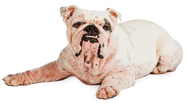
‘Scabies’ refers to infestation by the Sarcoptes scabiei mite, where the mite burrows through the skin, causing intense itching and irritation. The less common form of mange is demodectic mange, caused by an overgrowth of Demodex mites, which normally live in small numbers within dogs’ hair follicles. Localised or juvenile-onset disease is most often seen in young dogs, while generalised disease in an adult dog may be associated with an underlying illness or suppression of the immune system.
For more information, see our article Mange.
Nasal solar dermatitis
see Collie Nose
Pemphigus complex
Pemphigus is a complex of autoimmune diseases where the body’s immune system attacks its own skin, usually with no known cause. Pemphigus foliaceus, the most common autoimmune skin condition in dogs, is characterised by pustules, erosions and crusts on the skin’s surface. It typically begins in middle age, often occurring spontaneously in dogs with no prior history of skin disease though can also be drug-related.
Pigmentary abnormalities
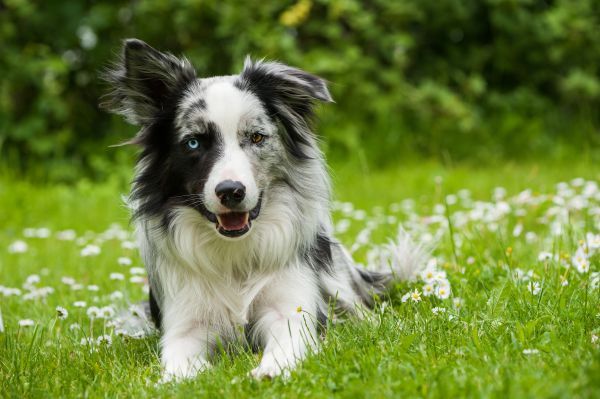
Skin and coat colour abnormalities may be acquired or hereditary, with abnormalities of the skin and coat colour often linked. Abnormally pale coat colours can be related to various health conditions, including neurological problems and deafness. Coats with dominant white or extreme piebaldism (spotted or blotched with black and white) may have related neurological problems or deafness in one or both ears.
A merle hair coat pattern, caused by the merle gene in dogs, is characterised by mottled patches of colour, blue or odd-coloured eyes and skin pigmentation issues. Deafness and other health issues may occur, particularly when two merles are bred together. Other pigmentary abnormalities include albinism, vitiligo and acquired aurotrichia (see these individual conditions above and below for more information).
Pruritus
Abnormal itching, called pruritus, occurs in many skin conditions and is often present because of secondary infections.
For more information, see Pruritus in dogs and cats.
Psoriasiform-lichenoid dermatosis
This genetic condition affects young Springer Spaniels, causing red symmetric abnormalities near the ears or inguinal region, comprising small lumps on the skin and solid elevated areas. These lumps can become increasingly thickened and spread to involve the face and trunk if left untreated.
Puppy impetigo (puppy pyoderma, juvenile pustular dermatitis)
Impetigo refers to small areas of infection found on the hairless area of the abdomen (belly). Puppy impetigo is a skin infection that usually occurs in puppies under one year of age and often develops in thinly haired areas such as the groin, underarms, chin and/or abdomen. Puppy impetigo is typically a localised area of infection. Pustules, small areas filled with pus, can be seen; these can break and form crusts or circular areas of scaling skin. Staphylococcus bacteria are usually present and thought to be a main cause of puppy impetigo. It is not life threatening and mild cases may resolve on their own.
Pyoderma (impetigo – see also puppy impetigo)
The term ‘pyoderma’ is derived from the Greek words for “pus” and “skin,” so this condition refers to a bacterial skin infection that exudes pus. Typically, lesions called papules or pustules, which may look like human pimples, form on the skin; these are usually red and raised, with a white pus-filled centre.
Pyoderma can also cause itching, redness, crusts, a rash, and/or hair loss at the site. There is often an underlying allergic dermatitis with pyoderma developing in the abrasions on the skin’s surface that occur from scratching. Pyoderma may also be referred to as impetigo, especially in young puppies.
Pyotraumatic dermatitis
Ringworm
see Dermatophytosis
Scabies
see Mite infestation (mange, acariasis, scabies)
Schnauzer comedo syndrome (Schnauzer comedone syndrome, Schnauzer back, Schnauzer Bumps)
In this genetic condition, Miniature Schnauzers develop bumps on the skin, generally affecting the area along the spine. Raised, black, crusty bumps (comedones) develop all along the back and neck. While normally not painful, they can become inflamed and sore if a secondary folliculitis and furunculosis occurs.
Sebaceous gland tumours and adenocarcinomas
Tumours of sebaceous glands are common in dogs. These glands secrete the oil known as sebum into the hair follicles and onto the skin. The tumours are often benign; however, sebaceous gland adenocarcinomas are a rare malignant form that occur in middle-aged or older dogs. They tend to grow rapidly, ulcerate and infiltrate into the deeper layers of the skin. The breeds most at risk are Cavalier King Charles Spaniels, Cocker Spaniels, and Scottish Terriers, Cairn Terriers, and West Highland White Terriers. Male dogs may be predisposed.
Expert advice from Dr Felicia:
Benign sebaceous gland tumours tend to behave predictably with little change. In some dogs they can get very large and irritating, and removal can help alleviate discomfort.
Seborrhoea, seborrheic dermatitis
This is a skin disorder in which the sebaceous (oil) glands of the skin produce an excessive amount of sebum. Abnormal turnover of skin cells (keratinocytes) into dead scale (keratin) is also involved, causing a build-up of keratin on the surface of the skin.
There are two types of seborrhoea: seborrhoea sicca (dry seborrhoea), which shows dry scaliness only, and seborrhoea oleosa (oily seborrhoea), in which the scales are accompanied by an oily coat, with a combination of these not uncommon. Primary seborrhoea is an inherited keratinisation disorder, while secondary seborrhoea is far more common, and results from disease processes such as allergies, parasites, nutritional disorders, and endocrine (hormonal) disorders such as hypothyroidism.
Bow Wow Meow’s Nose-to-Tail Cover has no sub-limits and up to $30,000 annual limit, giving you flexibility to use the full annual limit on your dog’s eligible skin condition management and treatment, if required, in a given year⑅.
Get a quick quote
Skin tags, hamartomas
These are distinctive, benign, stalk-like skin growths that usually occur on older dogs. Skin tags are common in dogs, may be single or multiple, and can develop in any breed, although large breeds may be more susceptible. Although unsightly, they are usually harmless. Dogs that develop one skin tag are likely to develop others.
Skin tumours
Tumours are abnormal growths of cells. Tumours affecting the skin or the tissue just under the skin are the most commonly seen tumours in dogs. They usually appear as small lumps or bumps, but they also can occur as blood blisters or non-healing ulcers.
The various layers and components of skin can develop distinctive tumours. While many skin tumours are benign, malignant (cancerous) tumours can occur. Benign tumours are not invasive and do not spread to other areas of the body, while malignant tumours can invade surrounding tissue and spread to distant organs.
Benign skin tumours include sebaceous adenomas, lipomas, histiocytomas, skin tags, fibromas and basal cell tumours. Malignant skin tumours include mast cell tumours, basal cell carcinomas, liposarcomas, squamous cell carcinomas and malignant melanomas.
Skin tumours can in some cases be associated with an underlying cancer occurring elsewhere in the body. For example, German Shepherds can develop nodular dermatofibrosis, where multiple nodules appear on the skin, generally on the head and legs. In most affected dogs, the condition is associated with cancer of the kidneys (renal cystadenocarcinoma), or of the uterus in females that have not been spayed.
Expert advice from Dr Felicia:
New skin and subcutaneous tumours should be mentioned and checked by the vet. Examination under the microscope is sometimes adequate for a diagnosis, but in other cases where there are abnormal cell populations, a small punch biopsy or excisional biopsy where the whole mass is removed for evaluation by a pathologist may be recommended.
Solar keratosis
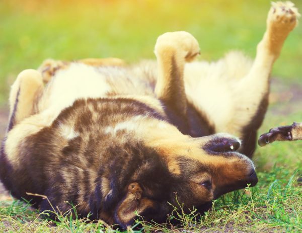
Solar keratosis is thickened and discoloured skin that is usually caused by prolonged and repeated sun damage. Because many animals sun themselves lying on their backs, it typically develops on the underside of dogs where the poorly haired skin offers minimal protection from ultraviolet radiation. Solar keratosis can progress to invasive squamous cell carcinoma.
Tumours
see skin tumours
Vitiligo
This is an uncommon but usually harmless condition of depigmentation, where the skin loses its natural pigment over time, causing patches of fading or white colour. Vitiligo is hereditary but is not noticeable at birth, with the onset in young adulthood. Affected dogs typically develop bleached splotches of skin on the face, especially the bridge of the muzzle or around the eyes, and occasionally also affecting the hair coat and claws. It affects certain breeds, especially Dachshunds, Dobermanns, German Shepherds and Rottweilers.
Warts
Signs and symptoms of skin conditions in dogs
There is a very broad range of symptoms relating to hair coat and skin conditions in dogs. These commonly include itching, redness, sores, change in smell and hair loss. Symptoms can occur throughout the skin or hair coat or in certain localised areas.
Other common symptoms of a skin or hair coat condition include:
- Dry, rough patches of flaking skin
- Excessive scaling, either in fine particles (dandruff) or sheets (coarse scale)
- Scales on face and paw pads
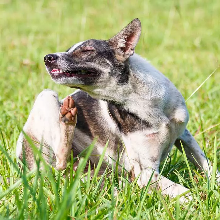
- Excessive itchiness, evident by scratching, rubbing, rolling, licking, chewing, head shaking, or scooting
- Hot spots, or localised areas of inflammation that your dog keeps scratching or biting
- Greasy accumulation of skin cells on the skin’s surface
- Moist, red sores under the hair coat
- Swelling or ulcerated skin
- Pimple-like pustules on the skin
- Seeping of blood or pus from irritated area
- Scabs, crusted skin
- Uncharacteristic hair loss, bald patches
- Uncharacteristic bad odour
- Infected hair follicles
- Hair follicles blocked up with oil and skin cells
Many of these symptoms can have multiple different causes, ranging from nutritional deficiencies, to endocrine disease or allergies.
When should you see a vet about your dog’s skin?
Arrange a veterinary appointment if your dog has persistent or recurring itching, excessive licking or chewing, unexplained hair loss, redness, scaling, a strong odour, sores, discharge or a lump that is new or changing. Early assessment can reduce self-trauma and secondary infection and may help identify an underlying allergy, parasite infestation or systemic illness.
Seek prompt veterinary care if the skin is very painful, the redness or swelling is spreading rapidly, there is bleeding or pus, your dog appears unwell or stops eating, or a lump is growing quickly or has become ulcerated.
Facial swelling, widespread hives or difficulty breathing may indicate a serious allergic reaction and require emergency veterinary attention.
What causes skin and coat conditions in dogs?
There are many possible underlying causes that can affect the appearance of a dog’s skin and coat, particularly its shine, texture and shedding. Some of the more common examples of potential systemic, underlying disease causing skin issues include hormone or metabolic disorders, parasites and cancer. Additionally, conditions affecting mobility, such as arthritis or obesity, can lead to skin problems such as dandruff or matting, if the dog is unable to groom itself properly.
The many and various underlying causes of hair coat and skin conditions in dogs may be broadly categorised as follows:
- External triggers
- Parasitic, bacterial, fungal and yeast infections
- Immune-mediated skin diseases
- Systemic diseases; hormonal (endocrine) and metabolic diseases or imbalances
- Hereditary conditions
- Nutritional deficiencies
- Contributing factors
External triggers
Skin inflammation and skin infections can occur in response to external stimuli – i.e. exposure to triggers in the environment or accidents that affect the skin. Triggers that can set off skin inflammation in dogs include external irritants/foreign bodies (e.g. chemicals, thorns, burrs), allergens (e.g. grass, dust mites, foods), burns, and trauma to the skin (e.g. a cut or dog bite).
Parasitic, bacterial, fungal and yeast infections
One of the most prevalent causes of skin conditions in dogs is external parasites (e.g. fleas, ticks, lice, mites). Depending on the geographical location, fleas can be extremely common in dogs, some of whom develop an allergic reaction in response to a flea bite, resulting in a flea allergy dermatitis.
Mites are another frequent underlying cause of skin disease in dogs, particularly Sarcoptes scabiei which cause scabies (sarcoptic mange) and less frequently, Demodex canis, which causes demodectic mange.
Underlying fungal or yeast infections can also lead to skin conditions, for example, ringworm (fungal) and Malassezia dermatitis (yeast). These infections can also occur secondary to a primary disease process compromising the skin barrier.
Bacterial skin infections are also widespread in dogs; however, they are typically a secondary reaction to another underlying condition such as parasites or allergies.
Abnormal immune reactions / immune-mediated skin diseases
Over-active, or hyper-defensive, immune responses include the body’s immune system attacking its own skin, leading to autoimmune skin disorders such as the pemphigus complex, systemic lupus erythematosus (SLE), discoid lupus, and vasculitis.
Expert advice from Dr Felicia:
Immune-mediated diseases (excluding allergies) in dogs are thankfully relatively uncommon but tend to require some additional diagnostics and a longer process to diagnose compared to infections/parasite infestations.
Systemic diseases
A systemic disease is one that affects several organs and tissues, or even the body as a whole. Systemic illnesses include endocrine disorders and hormonal imbalances, as well as metabolic diseases such as diabetes mellitus, renal disease, and liver disease.
Some systemic diseases can become symptomatic as a skin disorder; in fact, the skin is often a window to systemic disease (i.e., the skin can be the first part of the body where signs of systemic illness are noted). Immunosuppression leads to increased susceptibility to mange and recurrent bacterial and/or fungal skin infections.
Skin and hair coat changes are a common manifestation of hormonal diseases such as hypothyroidism, hyperadrenocorticism (Cushing’s disease), and sex hormone abnormalities, including tumours of the ovaries or testicles.
Early recognition of skin changes as clinical markers of systemic disease is important in expediting diagnosis and treatment of the underlying disorder.

Hereditary conditions
The term “genodermatoses” applies to skin conditions of genetic origin. Dogs can inherit several different kinds of skin disorders; these may be present at birth or may appear later. Many of these diseases are inherited in a recessive pattern, which means affected puppies can be born to seemingly normal parents.
Genetic skin disorders can occur in a wide range of dog breeds, although some manifest with greater frequency in certain breeds. Most of these conditions are uncommon to rare.
Some of the breeds more prone to particular hereditary or congenital skin conditions include:
- Standard Poodles – sebaceous adenitis, which affects the oil glands and can cause hair loss and a moth-eaten appearance.
- British Bulldogs – histiocytomas, or skin tumours that appear as a lump on the skin.
- Rhodesian Ridgebacks – Dermoid cysts, skin pockets into which dander, hair, oil, and other debris accumulate.
- Irish Terriers – Familial footpad hyperkeratosis, increased thickness of the hard, outer layer of the skin.
- English Springer Spaniels – Psoriasiform-lichenoid dermatosis, small red lumps on the skin and solid elevated areas near the ears or groin.
- Rottweilers – Vitiligo, bleached splotches of skin on the face, especially the bridge of the muzzle or around the eyes.
- Miniature Schnauzers – Acquired aurotrichia, where the hair along the middle of the back above the spine changes from the normal black or grey to golden.
- Chinese Shar Peis – Cutaneous mucinosis, where the skin exhibits pronounced folding and blisters.
- Cocker Spaniels – primary disorders of keratinisation
- Terriers – Atopic dermatitis, where exposure to an environmental allergen results in an inflammatory skin response.
Nutritional deficiencies
The nutrients that most commonly affect the quality of skin and coat are protein and fatty acids. Skin and hair, because of their constant renewal, utilise a significant portion of dietary protein; therefore, when there is inadequate or poor-quality protein in the diet, the coat and skin are among the first areas of the body to be affected. The outer layer of the skin, known as the stratum corneum, utilises essential fatty acids to prevent excessive water loss in the skin.
Also affecting the skin and hair coat are deficiencies of vitamin A and zinc, as well as general malnutrition. When the diet is inadequate to meet the dog’s dietary needs, it will have a dull, dry hair coat and will often shed excessively.
Expert advice from Dr Felicia:
Dogs with nutritional deficiencies causing dryness of the coat can also often benefit from topical therapies in addition to diet change.
Additional contributing factors to skin and hair coat disorders
- Age – distinct age predilections are seen in many skin diseases, e.g., demodicosis and dermatophytosis in paediatric animals; atopic dermatitis in 1–3 yr olds.
- Pruritus – conditions that begin with pruritus may lead to self-trauma and subsequent development of secondary skin lesions (e.g., alopecia, seborrhoea) or infections (bacterial or yeast pyoderma).
- Seasonal change – correlating with fleas, allergic skin disease, or weather-related diseases.
- Stress or excessive boredom
- Frequency of bathing – excessive bathing and wetting of the skin can predispose to skin disease.
- Contact with other animals – contact with an infested animal can mean dogs will also be infested with fleas, scabies, cheyletiellosis, or dermatophytosis.
- Environmental conditions – housing conditions can influence the development of certain skin diseases, e.g. frequent access to sunlight or excessive moisture in the home.
How are hair coat and skin conditions in dogs diagnosed?
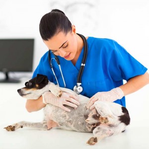
It is strongly recommended that hair coat and skin conditions in dogs are evaluated by a veterinarian to determine the cause and establish the correct diagnosis and the most effective form of treatment.
Because there are numerous dog skin conditions with similar symptoms, the underlying cause may not be immediately evident. In this case, the veterinarian will take a stepwise approach to investigating the possible disorders that could be causing the symptoms. This may be by use of treatment trials, dietary trials, blood tests or biopsies.
A physical examination, as well as a general history of the dog’s health and a detailed dermatological history will be required. Based on the findings, various tests and/or procedures may be required. Several consultations may be necessary for an accurate diagnosis to be obtained.
Physical examination
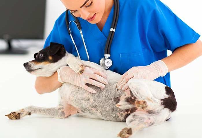
A complete physical examination should always be performed. Many skin diseases are indicators of systemic diseases (i.e. diseases that affect other parts of the body or the entire body). A good dermatologic examination entails a very close inspection of the entire hair coat and skin under strong lighting. It is important to thoroughly examine the dog’s all over so as not to miss any lesions and to closely palpate the skin to not miss lumps on haired areas.
History
A good history is important, because many skin diseases that look similar are differentiated based on interpreting clinical signs in conjunction with historical patterns. General information about prior illnesses, vaccinations, housing, feeding practices, changes in attitude and food consumption, elimination practices, exposure to other animals, and travel within the past 6 to 12 months should be provided.
A detailed dermatologic history is essential to interpret the physical examination findings and choose appropriate diagnostic tests. The following dermatological information should be provided:
- Onset and progression of the disease or primary complaint
- Length of time the condition has been present
- Age at which the condition started
- Presence and severity of pruritus (severe itching, characterised by behaviours including licking, rubbing, scratching, or chewing)
- Type and progression of lesions observed by the owner
- Evidence of seasonality
- Area on the body the problem was first noticed
- Any previous treatments for skin conditions and the responses to such
- Presence of fleas, ticks, or mites and use of parasite prevention
Expert advice from Dr Felicia:
Frequent recurrence of skin disease in patients should prompt a revision of the management plan or further evaluation of the initial issue.
Based on the physical examination findings and history, the veterinarian may perform several tests, which could include:
- A complete blood count, biochemistry profile, urinalysis and faecal testing to evaluate general health; however, in most dermatologic cases, these tests do not help to make a definitive diagnosis. If the dog has signs of systemic illness, these tests may help to identify the cause, while in dogs with recurrent infections, they may identify an underlying subclinical disease.
- Skin scrapings, i.e. collecting material from the skin’s surface (superficial) or within the hair follicle (deep). This material can be examined under a microscope to detect the presence of mites or other abnormalities.
- Cytologic examination of the skin. This can be taken by a cotton swab, sticky tape or by directly placing a slide on moist skin to evaluate the cellular population and any micro-organisms such as bacteria or yeast that may be present.
- Combing of the hair coat or “flea combing,” to collect large amounts of skin debris and trap parasites such as fleas, ticks, lice, and some mites.
- Bacterial and fungal cultures
- Skin biopsies in cases that appear severe, unusual, or do not respond to appropriate therapy
- Serum immune tests, if an allergy is suspected
- Intradermal allergy testing, in cases of severe and chronic allergic symptoms
- Food elimination trial, if a food ingredient is suspected
- Hormonal assays
Outlook and long-term management
The outlook depends on the cause and severity of the skin condition. Parasites, minor infections and some localised skin problems may resolve completely with appropriate treatment. Allergic, hormonal, immune-mediated and inherited conditions are more likely to require ongoing monitoring or lifelong management.
Recurring symptoms do not necessarily mean that treatment has failed. They may indicate a flare-up, secondary infection, a new trigger or a need to adjust the management plan. Following the prescribed treatment, parasite prevention, dietary trial and follow-up schedule gives the best chance of keeping the dog comfortable and protecting the skin barrier.
Expert advice from Dr Felicia:
For some patients management of their skin issues may require lifelong therapies, and regular vet visits. Having a management plan as well as a plan in place for ‘flares’ is helpful for keeping your pet’s coat healthy and comfortable long term.
Treatment and management of dog skin conditions
Successful treatment of hair coat and skin disorders requires definitive diagnosis of the specific condition. If the dog’s skin or coat problem is caused by an underlying health issue, the general health of the skin and the quality of the hair should improve dramatically when the illness is brought under control with appropriate treatment.
In many cases, a multimodal treatment approach will be prescribed encompassing two or more types of treatment. These may include:
Topical treatment
Many treatments for skin diseases are applied topically, i.e. directly to the surface of the skin. This may be the only treatment, or it may be prescribed in conjunction with systemic treatment (medications taken by mouth or injected and distributed throughout the body). Clipping or shaving the hair coat may be recommended to allow the topical therapy to reach the skin more easily, making treatment much more effective.
Topical treatment products can be in the form of ointments, creams, gels, shampoos and sprays. The vet may prescribe, for example, antibiotic ointment, corticosteroid cream, medicated shampoo and/or topical insecticide. It is essential to closely follow instructions for all topical medications; they should be used in the appropriate amounts for adequate effect. It is also important to discuss strategies to reduce the likelihood of dogs licking and ingesting topical medications.
Medicated shampoos are frequently used for specific skin conditions, including antimicrobial and antiseborrheic products. Regular application of a medicated shampoo is very important; often as much as 2 to 3 times per week initially. The medicated shampoo is applied evenly to the coat for the recommended contact time, then thoroughly rinsed off.
Expert advice from Dr Felicia:
Often some distraction techniques will be required to prevent dogs from simply licking off their topical treatments. Distractions may involve taking them for a walk or feeding a chewy treat that requires their active concentration to consume.
Medication / drug therapy
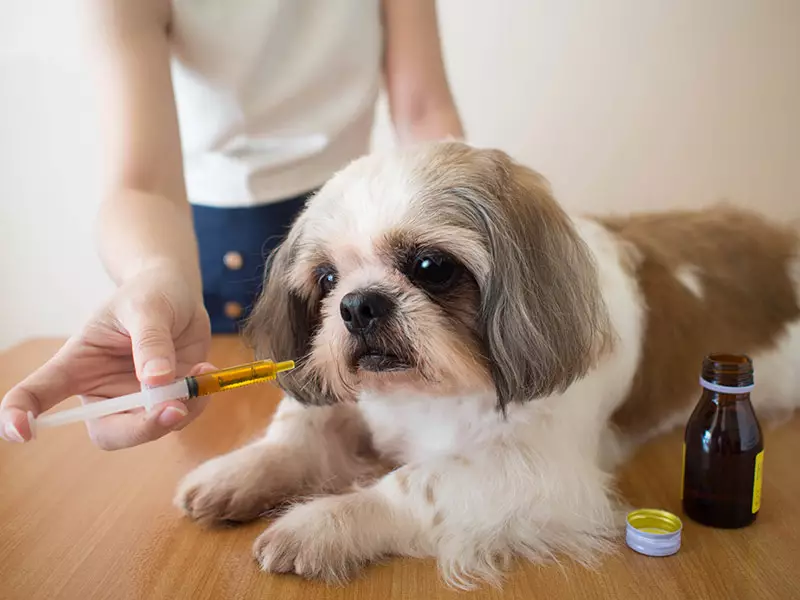
Systemic drugs may be prescribed by the veterinarian to treat some skin and haircoat disorders. These include antibiotics, antifungals, anti-parasitics, hormone therapy, antihistamines, corticosteroids and other anti-inflammatory drugs. These should always be administered according to the vet’s instructions.
Dietary modification and supplementation
To maintain the skin and hair in a healthy state, a properly balanced diet containing high quality digestible proteins, carbohydrates, fats, minerals and vitamins, as well as enough calories to meet the animal’s energy needs, is required. Therefore, treatment may include dietary changes and/or dietary supplementation.
For nutritional deficiencies of specific vitamins, such as vitamin A, vitamin E or zinc, supplements will be prescribed. For more broad nutritional deficiencies caused by general malnutrition, a balanced diet and a supplement of essential fatty acids may be prescribed. Some dogs are more sensitive than others, but all dogs benefit greatly from a healthy diet that does not contain an abundance of filler or artificial ingredients.
Skin tumour treatment
The appropriate treatment for a skin lump depends on its type, size, location, behaviour and whether it is causing discomfort. Some benign growths can be monitored once a vet has confirmed the diagnosis, while others may need to be removed because they are growing, ulcerated, infected or interfering with the dog’s normal activities.
A lump’s appearance alone cannot reliably show whether it is benign or malignant. Your vet may recommend collecting cells with a fine-needle aspirate or taking a biopsy before deciding whether monitoring, surgical removal or another treatment is appropriate.
Expert advice from Dr Felicia:
Whether or not tumours should be removed will depend on their behaviour (benign/malignant), location and if it is causing distress to the pet, as some dogs will lick or chew at their lumps which can cause pain and infection.
Treating the underlying condition
In most cases, the skin condition will improve once the underlying condition has been identified and properly treated. This may entail specific medication to treat a systemic disorder such as hypothyroidism or Cushing’s disease. Different types of treatment may be required depending on the underlying cause of the condition, for example:
Treatment of alopecia
Depending on its underlying cause, treatment may entail:
- Flea control
- Antibiotics, for skin infections
- Various treatments of skin allergies
- Medication for hormonal conditions (long-term treatment)
- Treatment for joint pain resulting from over-grooming
Treatment of scaly skin
Depending on its underlying cause, treatment may entail:
- Topical products, such as medicated shampoos (salicylic acid and benzoyl peroxide aid in cell turnover), or dips
- Targeted medication to prevent and treat parasites or infections
- Antibiotics for bacterial skin infections
- Antifungal drugs for fungal infections
- Stronger antiparasitic drugs if topical treatments are not strong enough
How can you support healthy skin and coat?
- Groom your dog regularly to remove loose hair, prevent matting and help you notice lumps, parasites or irritated areas early. The appropriate grooming frequency depends on the breed and coat type.
- Use effective parasite prevention suitable for your dog’s age, health, lifestyle and location.
- Feed a complete and balanced diet appropriate for your dog’s life stage.
- Bathe your dog at a frequency suitable for its coat and skin. Use medicated or hypoallergenic products only as directed by your vet.
- Keep skin folds clean and dry where appropriate, particularly in dogs prone to fold dermatitis.
- Keep nails at a comfortable length to reduce damage caused by scratching.
- Seek veterinary advice if itching, redness, hair loss, sores or an unusual odour persist or recur.
- Protect vulnerable, lightly pigmented skin from excessive sunlight. Use only a pet-safe sunscreen recommended by your vet.
- Do not clip or shave the coat solely for summer cooling without first considering the dog’s coat type and seeking appropriate advice.
Expert advice from Dr Felicia:
Ultimately some dogs will be more prone than others to skin disease. Breed will play a part, but ensuring your dog’s coat is kept clean and at an appropriate length will help support skin health. Catching skin disease early will often help to speed recovery.
In a nutshell…
Skin and coat changes in dogs can be caused by anything from parasites and minor infections to allergies, hormonal disease, inherited disorders and skin tumours. Common warning signs include persistent itching, licking, hair loss, redness, scaling, sores, odour and new or changing lumps.
Because several conditions can produce almost identical symptoms, an accurate diagnosis is important before treatment begins. Many skin problems can be treated successfully, while chronic conditions can often be controlled with an individual management plan and regular veterinary follow-up.
Bow Wow Meow Pet Insurance can help protect you and your dog should an unexpected trip to the vet occur.
-
Find out more about our dog insurance options
-
Get an online pet insurance quote
Bow Wow Meow is proud to have been awarded winner of Canstar’s ‘Most Satisfied Customers’ Award in the Pet Insurance category for both 2024 and 2025!
Bow Wow Meow is proud to be the only Pet Insurer that has been awarded Product Review’s Top Rated Pet Insurance every year from 2018 to 2026! This is based on 3,193 independent customer reviews (as at 09/04/2026), with an overall rating of 4.5 stars.

We also have the following ratings on other review platforms:
Google Review rating of 4.5 stars (based on 1,358 reviews)
Trust Pilot rating of 4.6 stars (based on 555 reviews)
Bow Wow Meow has been chosen as Winner of 4 categories in the 2026 Finder Pet Insurance Customer Satisfaction Awards!




– Most Trusted Pet Insurance
– Easiest to Claim Pet Insurance
– Most Recommended Pet Insurance
– Legendary Service Pet Insurance
We also received Highly Commended Awards for Loved Brand Pet Insurance and Best Value Pet Insurance.
Bow Wow Meow is an Australian-owned and operated pet insurance provider. Our focus is purely on pets, and we are proud to have helped provide peace of mind to over 200,000 Aussie pet owners since 2008.
Sources
“Diagnosis of Skin Diseases in Small Animals”, MSD Veterinary Manual, https://www.msdvetmanual.com/integumentary-system/integumentary-system-introduction/diagnosis-of-skin-diseases-in-small-animals. Accessed 4 August 2026.
“Dermatological Problems in Animals”, MSD Veterinary Manual, https://www.msdvetmanual.com/integumentary-system/integumentary-system-introduction/dermatological-problems-in-animals. Accessed 4 August 2026.
“Structure of the Skin in Dogs”, MSD Veterinary Manual, https://www.msdvetmanual.com/dog-owners/skin-disorders-of-dogs/structure-of-the-skin-in-dogs. Accessed 4 August 2026.
“2023 AAHA Management of Allergic Skin Diseases in Dogs and Cats Guidelines”, American Animal Hospital Association, https://www.aaha.org/resources/2023-aaha-management-of-allergic-skin-diseases-in-dogs-and-cats-guidelines/. Accessed 4 August 2026.
“Atopic Dermatitis in Dogs”, MSD Veterinary Manual, https://www.msdvetmanual.com/integumentary-system/atopic-skin-conditions-in-dogs-and-cats/atopic-dermatitis-in-dogs. Accessed 4 August 2026.
“Pyoderma in Dogs and Cats”, MSD Veterinary Manual, https://www.msdvetmanual.com/integumentary-system/pyoderma/pyoderma-in-dogs-and-cats. Accessed 4 August 2026.
“Mite Infestation (Mange, Acariasis, Scabies) in Dogs”, MSD Veterinary Manual, https://www.msdvetmanual.com/dog-owners/skin-disorders-of-dogs/mite-infestation-mange-acariasis-scabies-in-dogs. Accessed 4 August 2026.
“Dermatophytosis in Dogs and Cats”, MSD Veterinary Manual, https://www.msdvetmanual.com/integumentary-system/dermatophytosis/dermatophytosis-in-dogs-and-cats. Accessed 4 August 2026.
“Tumors of the Skin in Dogs”, MSD Veterinary Manual, https://www.msdvetmanual.com/dog-owners/skin-disorders-of-dogs/tumors-of-the-skin-in-dogs. Accessed 4 August 2026.
“Nutritional Requirements of Small Animals”, MSD Veterinary Manual, https://www.msdvetmanual.com/management-and-nutrition/nutrition-small-animals/nutritional-requirements-of-small-animals. Accessed 4 August 2026.
“Frequently Asked Questions and Myths”, World Small Animal Veterinary Association Global Nutrition Committee, https://wsava.org/wp-content/uploads/2020/01/Frequently-Asked-Questions-and-Myths.pdf. Accessed 4 August 2026.